beginning
In this article, I will explain the “plantar aponeurosis” in detail. The plantar aponeurosis is one of the thick membranes on the back of the foot, acts as a cushion during walking and running, and has an important role in supporting the arch of the foot. This article explains in detail the structure and function of plantar aponeurosis, common disorders such as plantar aponeurosis, and prevention measures and treatments. In order to maintain foot health, plantar aponeurosis care is essential, so it is important to learn its importance and proper care methods. Please use this article to deepen your understanding of the body.
Click here to watch a video about plantar fascia (plantar fascia)
teamLab Body Pro Free Download
A 3D anatomy app that shows all the structures of the human body
Download teamLab Body Pro here!

What is plantar aponeurosis
The plantar aponeurosis is an important structure that supports the arch of the foot. It absorbs shock applied during walking or driving, and plays a role in maintaining foot stability. A healthy plantar aponeurosis is an essential part of comfortable walking and sports activities.
How to read plantar aponeurosis
The correct way to pronounce plantar aponeurosis is “soku teikemmaku.” An aponeurosis (aponeurosis) refers to a membranous tissue where tendons are gathered together.
Characteristics of plantar aponeurosis
The plantar aponeurosis is strong yet flexible to a certain extent. Due to this property, it is possible to support body weight and hold the arch of the foot.
Location and location of plantar aponeurosis

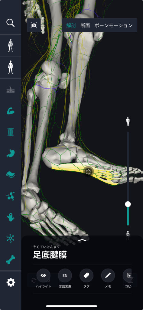
If you use a human anatomy diagram, you can see that the plantar aponeurosis is located inside the foot. Specifically, it extends from the heel bone (heel bone) to the front of the foot and is attached near the base of the toe.
How to remember plantar aponeurosis
The plantar aponeurosis is the “bottom - bottom” part, and it's a good idea to remember it as the “tendon membrane” at the “bottom” of the foot. Also, one method is to memorize with the image of supporting the arch of the foot.
English and Latin for plantar aponeurosis
Plantar aponeurosis in English*” Plantar Fascia”,It is expressed as “Fascia Plantaris” * in Latin. This knowledge will be useful when reading medical literature.
Plantar aponeurosis trivia
Plantar aponeurosis is a disease in which the plantar aponeurosis becomes inflamed. It is a disease that often occurs mainly due to prolonged standing work or wearing inappropriate shoes.
Tissues associated with plantar aponeurosis: characteristics of foot arches
The arch of the foot refers to the part of the foot that is not in contact with the ground. This arch is responsible for shock absorption, weight distribution, and balance when walking. The plantar aponeurosis is a layer of strong tendon that extends from the heel bone to the toe bone at the front of the foot. This aponeurosis supports the arch of the foot and absorbs the impact applied with each step, reducing fatigue when standing or walking for a long time.
There are two main types of foot arches. One is an inner longitudinal arch, and the other is an outer longitudinal arch. The medial longitudinal arch is tall and is the main arch that supports the inside of the foot. The lateral longitudinal arch is relatively low and is located on the outside of the foot. A healthy plantar aponeurosis is essential for these arches to function properly.
Tissues associated with plantar aponeurosis: location and position of foot arches
The arch of the foot is located on the bottom of the foot. The medial longitudinal arch is formed from the heel bone to the base of the big toe (thumb). This creates an inner elevation of the foot and provides stability when walking. The lateral longitudinal arch extends from the heel bone towards the little toe (little finger) and is lower than the medial longitudinal arch.
The plantar aponeurosis is the foundation of this arch structure and plays an important role in maintaining the shape of the foot and absorbing shock when walking. Inflammation or damage to the plantar aponeurosis can cause arch dysfunction and lead to a disease called plantar fasciitis.
Tissues associated with plantar aponeurosis: foot arch trivia
The structure of the arch of the foot can vary greatly from person to person. Some people have high arches (arches), while another group of people have so-called flat feet with few arches. This difference is influenced by genetic factors, weight, exercise habits, and even the type of shoes you wear.
Improper shoe selection and excessive exercise can put too much stress on the plantar aponeurosis and cause foot arch damage. Therefore, it is important to choose the right shoes for your foot arch type and do proper stretching before exercise.
Proper weight management, choosing the right shoes, and regular foot stretching are important for maintaining the health of the plantar aponeurosis and foot arch. With these measures, the arch of the foot and plantar aponeurosis will function properly and will be able to support our daily lives in a healthy way.
Plantar aponeurosis quiz and correct answers
Q. What part of the body is the plantar aponeurosis located?
1. soles
2. palms
3. shoulders
4. laps
A: 1. soles
As the name suggests, the plantar aponeurosis is located on the bottom of the foot. Let's understand it correctly and use it in our daily lives and sports activities.
summary
This time, I explained the location and location of “plantar aponeurosis”, how to remember it, and the English and Latin notation.
How was it?
I would be happy if reading this article deepened my understanding of anatomy.
Learning is a long, never-ending journey, but I sincerely wish you all the best. Let's continue to study together and work hard for the national exam!
Please look forward to the next blog.
Learn more with the anatomy app “TeamLabBody Pro”!
teamLabBody Pro is a “3D human anatomy application” that covers the entire body of the human body, including muscles, organs, nerves, bones and joints.
The human body is faithfully reproduced from CT and MRI data based on data from multiple subjects. Since medical book-level content supervised by physicians can be freely viewed from all angles, it can be used for various medical situations, such as explaining surgery to patients and studying anatomy for students.
If you want to see the parts introduced this time in more detail, please download the anatomy application “teamLabBody Pro.”
teamLab Body Pro Free Download
A 3D anatomy app that shows all the structures of the human body
Download teamLab Body Pro here!


