Superior oblique muscle: position, actions and trochlear nerve
Where is the superior oblique?
A well-bucket rope changes direction over a pulley. Did you know that the same device exists inside the orbit? The superior oblique is one of the six muscles that move the eyeball; its tendon passes through a small loop and doubles back, so the direction in which the muscle shortens is very different from that in which it pulls on the eyeball.
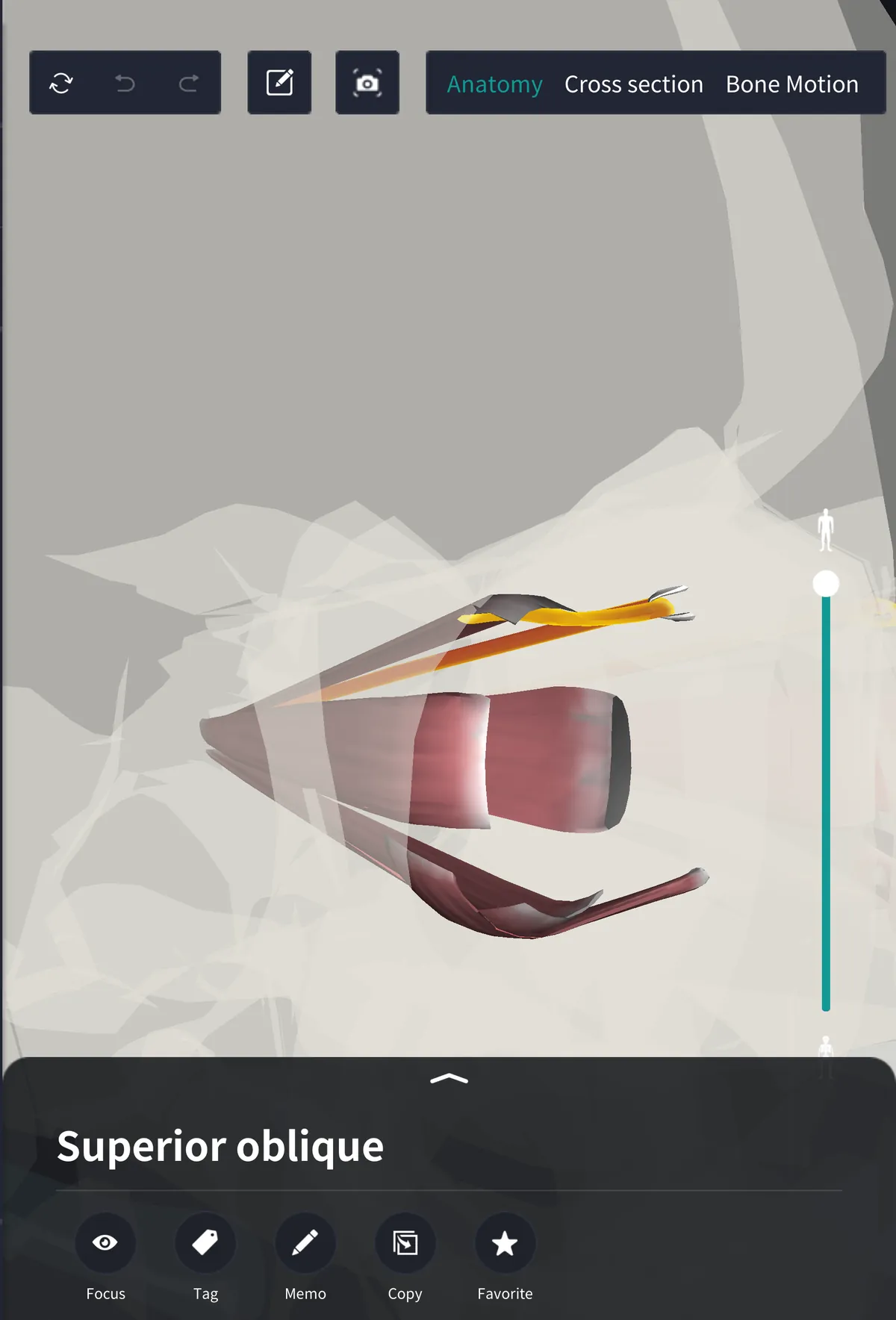
The view shows the orbit obliquely from the front, the muscles round the eyeball being seen through the skull. The slender coloured muscle is the superior oblique, running from the back forward along the junction of the inner wall and the roof of the orbit.
From origin to trochlea
It arises at the very back of the orbit, from the bone above and medial to the opening for the optic nerve, just outside the tendinous ring from which the four straight muscles arise. The long slender muscle passes forward along the upper inner angle of the orbit and becomes a rounded tendon short of the front.
The tendon passes through a loop. Attached to the bone at the upper inner part of the entrance to the orbit is a small ring of cartilage, the trochlea, deep to a point a little below the inner end of the eyebrow. The tendon runs through it and there changes direction.
From trochlea to eyeball
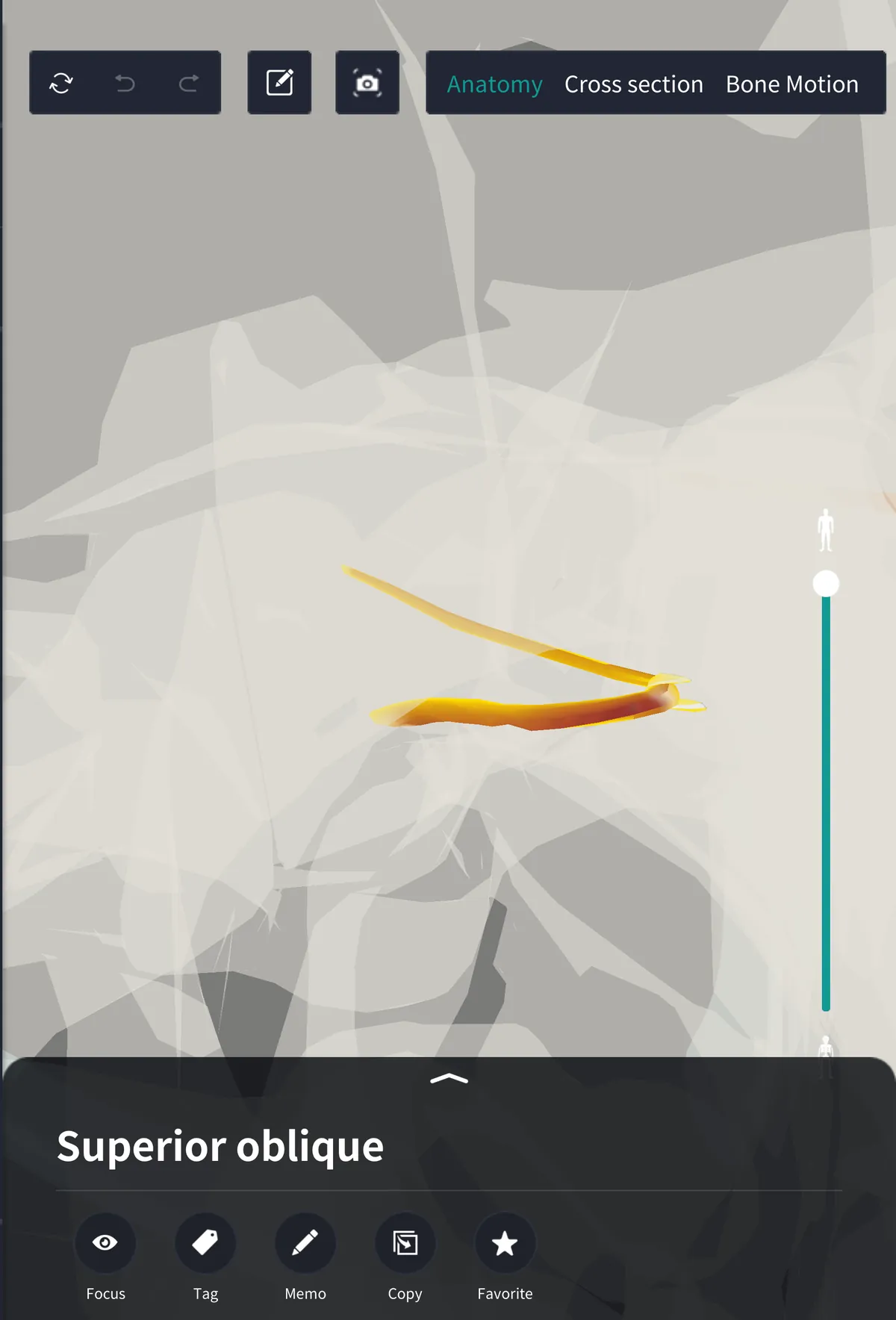
The view is taken obliquely from above. On leaving the trochlea the tendon doubles back: it passes backward, outward and a little downward, beneath the superior rectus covering the top of the eyeball, and attaches to the white of the eye (sclera) on the upper surface toward the back and outer side.
The attachment is on the back half of the eyeball, the reverse of the four recti, which attach to the front half. And since the tendon pulls from in front and medially, when the superior oblique contracts the upper outer back part of the eyeball is drawn toward the trochlea.
The superior oblique is the longest of the extraocular muscles. Its fleshy part extends only from the back of the orbit to the trochlea; the part beyond, doubled back, is all tendon, so only a slender tendon reaches the eyeball.
Action
It has three actions.
・Rotates the eyeball inward: tilts the top of the eyeball toward the nose, a twisting movement (intorsion)
・Turns the eye downward
・Turns the eye outward: weaker than the downward movement
Which is most marked depends on the direction of the eye. The tendon runs obliquely from in front and medially to behind and laterally. When the eye is turned toward the nose the line of sight comes into line with the tendon, and the superior oblique becomes a muscle that draws the eye straight down, as in reading or going downstairs, when one looks downward and inward. With the eye turned outward it is the reverse: the downward pull weakens and twisting becomes the chief effect.
In looking down two muscles work as a pair, the inferior rectus and the superior oblique. The inferior rectus acts chiefly when the eye is turned outward and the superior oblique when it is turned inward, so the eye can be directed downward in any position.
The opposite action belongs to the inferior oblique, which arises from the front of the floor of the orbit, winds backward and outward beneath the eyeball, rotates the top of the eyeball outward and turns the eye upward.
Nerve supply
The superior oblique is supplied by the trochlear nerve, the fourth cranial nerve, named from the pulley of this muscle. It supplies this one muscle only. It is the most slender of the cranial nerves and the only one to leave the back of the brain, and on entering the orbit it enters the muscle on its upper surface. Most of the other extraocular muscles are supplied by the oculomotor nerve.
Name and terms
Superior oblique means the upper slanting muscle; below the eyeball is the inferior oblique, which has no pulley. Trochlea is Latin for a pulley. Related terms: trochlear nerve, intorsion, depression of the eye, sclera, common tendinous ring.
See it in the app
The action of the superior oblique is notoriously hard to remember, because a muscle called superior turns the eye downward. Showing the superior oblique and the trochlea in the 3D anatomy app teamLabBody and looking straight down shows the tendon doubling back in front and medially and pulling the outer back part of the eyeball forward: as the back is drawn up and forward, the cornea turns down. The muscles in the model are seen through the skull, so their colour is paler than it is, and the slender tendon is hard to see without enlarging.
FAQ
- Where is the superior oblique?
- In the upper inner part of the orbit: it runs forward from the back, its tendon turns at the trochlea and it attaches to the upper back and outer part of the eyeball.
- What does the superior oblique do?
- It rotates the top of the eyeball inward and turns the eye downward and outward.
- Which nerve supplies the superior oblique?
- The trochlear nerve.
- Where is the trochlea?
- It is a small ring of cartilage attached to the bone at the upper inner part of the entrance to the orbit; the tendon of the superior oblique passes through it and changes direction.
Related articles
・Trochlear nerve: the fourth cranial nerve, supplying the superior oblique alone
・Superior and inferior rectus: the muscles that move the eye up and down
・Lateral and medial rectus: the straight muscles on the outer and inner sides, moving the eye from side to side
・Oculomotor nerve: the nerve of four of the extraocular muscles