
beginning
In this article, I will explain effective study methods, starting with knowledge of specialized parts in human anatomy.
In human anatomy, it is necessary not only to memorize the names of various organs, muscles, and bones, but also to remember where they are located in the body. Therefore, it is necessary to learn as efficiently as possible.
I hope you will deepen your understanding even a little by reading this article and using the app.
Now, I will explain the details about “rectus frontal/lateral rectus cephalus muscle” and how to study human anatomy.
teamLab Body Pro Free Download
A 3D anatomy app that shows all the structures of the human body
Download teamLab Body Pro here!

What is the rectus frontalus muscle and lateral rectus cephalus muscle?
In the anatomy application, you can view a selection of anatomy 3D models. In this model, there are various observation methods such as surfaces, cross-sections, and nervous systems. This time, I'll explain using an anatomy application.
About the rectus frontal muscle

The rectus frontalis muscle (frontalis) is a muscle located on the forehead of the face, attaches to the skin on the forehead, and has the function of opening the eyes wide and causing wrinkles on the forehead. This muscle plays an important role in the expression of emotions and movements that expand vision. The rectus frontal muscle works together with the occipital muscle as the “anteropharyngeal muscle” to support the movement of pulling the forehead up.
When the rectus frontal muscle contracts, it pulls the eyebrows upward and wrinkles the forehead. This allows us to express surprise, doubt, and focus. As part of facial muscles, it is controlled by the facial nerve (7th cranial nerve). In order to enhance facial expression, the rectus frontal muscle is used on a daily basis and plays an essential role in expressing emotions and reactions.
If the rectus frontal muscle is excessively tense over a long period of time, deep wrinkles remain on the forehead, and stress and fatigue may appear on the facial expression. Therefore, maintaining relaxation and flexibility of the rectus frontal muscle is important for healthy facial expression and facial care.
Study points
Anatomical understanding
The rectus frontalis muscle (frontalis) is a muscle that is part of the facial facial muscles and is located on the forehead. This muscle runs across the frontal region and extends over the upper part of the face. As a first step in learning, it is important to accurately understand where the rectus frontal muscle begins and stops. Together with the occipital muscle, the rectus frontal muscle functions as an overall “anteropharyngeal muscle (epicranius)” and plays a role in raising the forehead.
The starting part of the rectus frontal muscle directly affects the skin on the forehead through cooperation with the occipital muscles located in the back of the head. The stop is on the skin of the forehead. By contracting the rectus frontal muscle, you can raise your eyebrows and wrinkle your forehead. This muscle is very important, especially when it comes to expressing emotions and expanding one's field of vision.
Anatomically, the rectus frontal muscle is a large facial muscle and is controlled by the facial nerve (7th cranial nerve). When learning, it is important to be aware of the direction in which muscles run, relationships with surrounding muscle groups, and intersections with blood vessels and nerves. For visual learning, it is useful to use anatomical diagrams and 3D anatomy apps.
Understanding muscle function
To understand the function of the rectus frontal muscle, it is important to understand its movement and its impact on daily life and emotional expression. By creating wrinkles on the forehead, this muscle creates expressions of surprise, doubt, and concentration. It is also used to widen the eyes and expand the field of vision. The rectus frontal muscle is innervated by the facial nerve and plays an important role in expressing emotions.
When the rectus frontal muscle contracts, the eyebrows rise and wrinkles appear on the forehead. This helps with the movement of opening the eyes and expanding the field of vision. Also, when there is excessive tension or stress, the rectus frontal muscle is frequently used, and wrinkles on the forehead increase. Deep wrinkles may also form on the forehead when tension or stress builds up over a long period of time.
Practical applications
In order to practically apply the understanding of the rectus frontal muscle, facial facial muscle exercises and relaxation are effective. In particular, stretches and massages to relieve facial tension are recommended. Stretching the rectus frontal muscle involves putting a hand on the forehead, applying light pressure, and lifting the eyebrows upward. This movement relaxes the muscles and softens the overall facial expression.
In addition, exercises and facial expression training to balance facial muscles are also important. When the function of the rectus frontal muscle decreases, expression of facial expressions becomes poor, which may affect the transmission of emotions. Therefore, consciously strengthening facial muscles and increasing flexibility can help maintain healthy facial expressions.
About the lateral rectus cephalus muscle
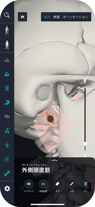
The lateral rectus muscle (lateral rectus muscle) is an important muscle that moves the eyeball outward, and is responsible for the abduction movement of the eyeball. This muscle starts from the outer wall of the eye socket and stops outside the eyeball. The lateral rectus cephalus muscle plays a role in moving the line of sight left and right, and is essential when looking at objects and for adjusting vision.
The lateral rectus cephalus muscle is innervated by the abductal nerve (sixth cranial nerve), and when this nerve is impaired, abductive movement of the eyeball may be impaired. Proper function of the lateral rectus cephalus muscle makes eye movements smoother and can reduce visual fatigue and discomfort.
When this muscle function declines, eye movement may be impaired and visual disharmony may occur. Therefore, maintaining the health of the lateral rectus cephalus muscle is important for maintaining eye motor function.
Study points
Anatomical understanding
The lateral rectus muscle (lateral rectus muscle) is an important muscle involved in eye movement, and is responsible for drawing the eyeball outward. Understanding the anatomy of this muscle begins by examining its starting and stopping parts. The lateral rectus cephalus muscle originates in the outer wall of the eye socket and stops outside the eyeball. This muscle is also called the abductor muscle because it moves the eyeball outward.
Anatomically, the lateral rectus cephalus muscle is very important for controlling eyeball movements. In order to advance learning, it is important to understand that the lateral rectus cephalus muscle is responsible for the abduction movement of the eyeball. You also need to know that this muscle is controlled by the adductor nerve (6th cranial nerve) other than the oculomotor nerve (3rd cranial nerve).
For visual learning, it is useful to use anatomical diagrams and 3D anatomy apps. In particular, it is important to accurately understand the cooperative relationship with other muscles involved in eye movement and the location of the lateral rectus cephalus muscle.
Understanding muscle function
In the functional understanding of the lateral rectus cephalus muscle, it is important to understand its role in the abduction movement of the eyeball. This muscle can direct the gaze from side to side by moving the eyeball outward. In particular, it is a necessary muscle for making left and right gaze and when looking at things.
If the lateral rectus cephalus muscle does not function properly, the abduction of the eyeball may be impaired, making it difficult to move the gaze from side to side. Such symptoms can cause eye movement disorders. Therefore, maintaining the function of the lateral rectus cephalus muscle is important for eye health and visual function.
Practical applications
In order to practically apply the understanding of the lateral rectus cephalus muscle, it is effective to perform eye movement exercises. In particular, in order to activate eye movement, it is effective to perform exercises to move the eyes left and right. By strengthening your eye muscles, you can prevent visual fatigue and eye strain.
In addition, rehabilitation and strength training related to the lateral rectus cephalus muscle are also effective. If visual dysfunction occurs, exercises to improve eye movement are recommended. This allows for smoother eye movement and improved visual comfort.
How to study human anatomy
I will explain specific study methods using human anatomy applications.
Check your past learning history and practice repeatedly
Here are the steps to check your anatomy learning history and practice iteratively effectively.
1. Check your learning history in the app
Reviewing your learning history with the application is an important step in effectively advancing anatomy learning. First, launch the app and go to the learning history section from the main menu. Many anatomy apps are designed to show your progress in the form of graphs and lists, so you can visually check which parts you've learned about and how much time you've spent.
By using this data, you can understand which areas you have strengths in and where you need to spend more time and effort. We also recommend using a dedicated tag or notebook function to mark areas you are particularly weak at or where you need to relearn. Regularly checking your learning history and looking back on past learning content will lead to efficient review and deepening understanding.
2.Make a plan for iterative learning
Making an efficient repetitive learning plan based on learning history is extremely effective in promoting knowledge retention. First, identify weak points and areas where you need to relearn. Next, arrange these study items into a weekly or monthly calendar and create a specific study schedule. By proceeding in a planned manner, you can learn each part evenly and avoid packing in a large amount of information at once.
Using a task management app or digital calendar to set study reminders is effective. Also, it's important to have the flexibility to regularly review progress and revise plans as needed. By having goals and proceeding with your studies in a planned manner, you can efficiently acquire anatomical knowledge.
3.Use 3D features to learn visually
By utilizing the 3D function, learning anatomy is easier to understand visually. The 3D model shows the structure of the human body three-dimensionally, and each part can be observed in detail. This makes it possible to intuitively grasp positional relationships between deep muscles and organs that are difficult to capture in a planar view. For example, you can learn even the smallest details by rotating specific muscles and bones and zooming in and out.
Also, there are many apps that have the function of displaying cross-sectional views of each part using a 3D model, which is useful for deepening understanding of internal structures. This diversity of visual information helps with memory retention and improves immediate responsiveness in tests and practice situations. By utilizing the 3D function and learning visually, you can learn anatomy knowledge more deeply and efficiently.
Use the memo function concretely

Make notes so you don't forget the things and points you've noticed while studying. The memo function can be used for different purposes, such as inputting text, saving images, and writing memos. Tag your notes to make them easier to review later.
Test your learning regularly in the form of quizzes
Regularly testing what you've learned in a quiz format is a very effective way to anchor your anatomy knowledge. Quiz-style tests help you objectively grasp your level of understanding and areas you lack while repeating knowledge.
For example, by using a learning app to conduct quizzes every specific period, you can reconfirm what you've learned and strengthen your memory. There are a wide range of quiz formats, such as multiple choice questions, fill-in-the-blank questions, and short answer questions, and each helps understanding from a different angle and develops the ability to utilize various types of knowledge.
Get feedback
If possible, get feedback from other learners and experts. It helps you find your own gaps in understanding and areas for improvement. You can also keep yourself motivated to learn by regularly testing yourself. Feeling a sense of accomplishment and progress increases motivation for continuous learning.
summary
This time, I explained how to study “rectus frontal/rectus lateralis muscle” using an application!
Thank you for reading this far.
I would be happy if reading this article helped you learn about anatomy.
Learning is a long, never-ending journey, but I sincerely wish you all the best. Let's continue to study together and work hard for the national exam!
Please look forward to the next blog.
teamLab Body Pro Free Download
A 3D anatomy app that shows all the structures of the human body
Download teamLab Body Pro here!