
Spinalis cervicis: origin, insertion and action
Introduction
This article first introduces specialist knowledge about one part of the body in human anatomy, then explains effective ways to study.
In human anatomy it is not enough to memorize the names of organs, muscles and bones; you also have to remember where in the body they are. That is why you need to learn as efficiently as possible.
I hope that reading this article and using the app will deepen your understanding, even a little.
This time, we cover the spinalis cervicis and how to study human anatomy.
Download teamLabBody Pro for free
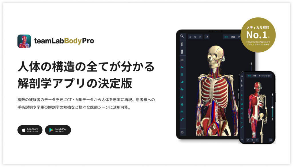
A 3D anatomy app that shows every structure of the human body
Download teamLabBody Pro here!
What is the spinalis cervicis?
In the anatomy app you can view a selection of anatomical 3D models. Each model can be viewed in several ways: the surface, cross-sections, the nervous system and more. This time we explain using the anatomy app.
About the spinalis cervicis

The spinalis cervicis is part of the spinalis, the innermost column of the erector spinae, and is a slender muscle running vertically near the midline at the back of the neck. It arises from the lower part of the nuchal ligament and the spinous processes of C7 to T2 and inserts on the spinous process of the axis (C2). It sometimes reaches the spinous processes of C3 and C4, and how well developed it is varies greatly between individuals.
When the left and right spinalis cervicis work together, they bend the neck backward (extension); when only one side works, it tilts the neck to the same side (lateral flexion). It is supplied by the posterior rami of the spinal nerves and, together with the deeper semispinalis cervicis and multifidus, supports the alignment of the cervical spine from behind. Because it is a thin muscle linking spinous process to spinous process, show the nuchal ligament and spinous processes in 3D to check its position.
Learning points
Anatomical understanding
The spinalis cervicis belongs to the erector spinae of the intrinsic back muscles. The erector spinae is arranged in three columns, from lateral to medial: iliocostalis, longissimus and spinalis. The spinalis cervicis is the neck part of the innermost column, the spinalis. Arising from spinous processes and inserting on spinous processes is the hallmark of the spinalis: the spinalis cervicis arises from the lower nuchal ligament and the spinous processes of C7 to T2, and inserts on the spinous process of the axis (sometimes C3 and C4). Just deep to it lie the semispinalis cervicis and multifidus (the transversospinalis group), which run obliquely from transverse processes to spinous processes; together they stabilize the spine and support neck movement.
As a first step, it is important to understand where the spinalis cervicis is and how it interacts with other muscle groups. Anatomical illustrations and 3D apps are very effective for checking the course of the muscle visually. Understanding its position relative to neighboring muscles (for example, the trapezius and semispinalis capitis) also makes the role of the spinalis cervicis clearer.
Understanding the muscle's function
When both sides of the spinalis cervicis work together, they extend the neck and bend it backward; when only one side works, it bends the neck laterally to the same side. It supports movement of the head and neck and is one of the muscles that maintain the alignment of the upper spine.
As you study, link the movements of the neck with the course of the spinalis cervicis. When you tilt your head back, the left and right muscles shorten together; when you tilt your neck to one side, the muscle on that side shortens. Display the course of the muscle in 3D and imagine which way it pulls in each movement to deepen your functional knowledge.
How to remember it: sort by the three columns of the erector spinae
The name spinalis cervicis combines one of the three columns of the erector spinae (from lateral to medial: iliocostalis, longissimus, spinalis) with a region (capitis, cervicis, thoracis, lumborum). Break it down as "the innermost column (spinalis)" plus "the neck part (cervicis)" and it becomes easier to tell apart from the longissimus cervicis and iliocostalis cervicis. As its name suggests, it runs from spinous process to spinous process, so display the nuchal ligament and the row of spinous processes at the midline of the back of the neck in 3D, and find it as the thin muscle running alongside them on each side.
How to study human anatomy
Here are concrete study methods using a human anatomy app.
Check your learning history and practice repeatedly
These are the steps for checking your anatomy learning history and reviewing effectively.
1. Check your learning history in the app
Checking your study history in the app is an important step for making effective progress in anatomy. First open the app and go to the history section from the main menu. Many anatomy apps show your progress in graphs or lists, so you can see which parts you have studied and how much time you spent on each.
With that data you can see where you are strong and where you need to invest more time and effort. It also helps to mark the parts you find difficult or need to relearn with tags or notes. Checking your history regularly and going back over what you have already studied leads to effective review and deeper understanding.
2. Make a review plan
Making an efficient review plan based on your learning history is extremely effective for consolidating knowledge. First identify your weak points and what you need to relearn. Then spread those topics over a weekly or monthly calendar and create a concrete study schedule. Working to a plan lets you cover every region evenly and prevents you from cramming too much information at once.
It is also effective to set study reminders with a task management app or a digital calendar. It is important to review your progress regularly and stay flexible enough to adjust the plan when necessary. Studying with goals and a plan lets you acquire anatomical knowledge efficiently.
3. Learn visually with the 3D functions
With the 3D functions, anatomy becomes much easier to understand visually. The 3D model shows the structure of the body in three dimensions and lets you observe each part in detail. You can grasp intuitively the positional relationships of deep muscles and organs that are hard to see in a flat figure. For example, you can rotate a specific muscle or bone and zoom in and out to study it down to the finest detail.
Many apps can also display cross-sections of each part on the 3D model, which helps you understand the internal structure. This variety of visual information helps you remember and improves your responsiveness in exams and in practice. Learning visually in 3D gives you deeper, more efficient anatomical knowledge.
Make use of the memo function
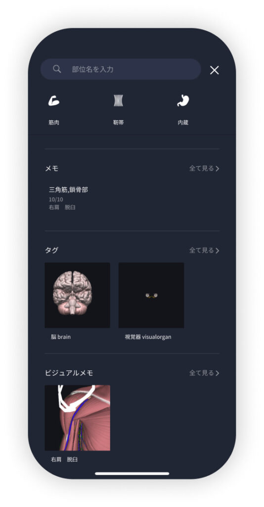
Make notes of what you notice and the key points while studying so that you do not forget them. The memo function lets you type text, save images or write by hand, depending on your needs. Tag your memos so that you can find them easily later.
Test what you have learned regularly in quiz form
Regularly testing what you have learned in quiz form is a very effective way to consolidate anatomical knowledge. Quizzes let you repeat the material while objectively checking how well you understand it and where the gaps are.
For example, taking quizzes in a study app at set intervals lets you recheck what you have learned and reinforce your memory. There are many quiz formats (multiple choice, fill in the blank, short answer), and each helps you understand from a different angle and use different kinds of knowledge.
Get feedback
If possible, get feedback from other learners or specialists. It helps you find what you have not understood and what you can improve. Testing yourself regularly also helps you stay motivated: feeling your achievements and progress makes you want to keep learning.
Summary
This time we explained how to study the "spinalis cervicis" with an app!
Thank you for reading this far.
I hope this article helps you with your study of anatomy.
Learning is a long road with no end, but I sincerely wish you success. Let's keep learning together and work toward the exam!
Look forward to the next article.
Download teamLabBody Pro for free
A 3D anatomy app that shows every structure of the human body
Download teamLabBody Pro here!


