Where is the hyoid bone? Location, greater and lesser horns, and attached muscles
Where is the hyoid bone?
Slide a finger from the tip of your chin down toward your Adam's apple. Just above it you will feel a hard bar running sideways: that is the hyoid. Pinch it gently from both sides with your thumb and index finger and swallow. The bone rises and drops back in a moment.
It lies at about the level of the third cervical vertebra, between the mandible and the thyroid cartilage of the larynx. Because it is not attached to the skull or the spine, it is easy to miss in a skeleton model, where it often hangs on a wire or sits in a separate bag.
Hyoid bone and thyroid cartilage
Which one is higher? The Adam's apple is the thyroid cartilage, and the hyoid sits about one finger's width above it. The thyrohyoid membrane stretches between them, so when the hyoid rises during swallowing, it lifts the larynx with it.
Parts of the hyoid
・Body: the square middle part at the front. Most suprahyoid and infrahyoid muscles attach to its front surface
・Greater horns: long, thin projections running backward and upward from the body. When you pinch the hyoid, your fingertips rest on them
・Lesser horns: small cone-shaped projections where the body meets the greater horns. The stylohyoid ligament from the styloid process of the temporal bone attaches here
The greater horns are joined to the body by cartilage in young people and often fuse to it by bone in middle age, though the timing varies from person to person.
Muscles attached to the hyoid
The muscles fall into two groups. The suprahyoid muscles lie above the bone: the digastric, stylohyoid, mylohyoid and geniohyoid. They raise the hyoid when you swallow, and when the hyoid is held still they pull the mandible down to open the mouth.
The infrahyoid muscles lie below it: the sternohyoid, omohyoid, sternothyroid and thyrohyoid. They pull the hyoid back down after swallowing. The sternothyroid is the exception that stops at the thyroid cartilage. The hyoglossus also arises from the body and greater horn and runs into the side of the tongue.
Innervation: the anterior belly of the digastric and the mylohyoid are supplied by the trigeminal nerve, the posterior belly and the stylohyoid by the facial nerve, the geniohyoid and thyrohyoid by C1 fibres travelling with the hypoglossal nerve, and the other infrahyoid muscles by the ansa cervicalis.
Name and Japanese term
The English name comes from Greek hyoeides, "shaped like the letter upsilon (υ)", after its U shape seen from above. Any muscle name ending in -hyoid attaches to it. In Japanese the bone is called 舌骨 (zekkotsu), literally "tongue bone".
See it in 3D
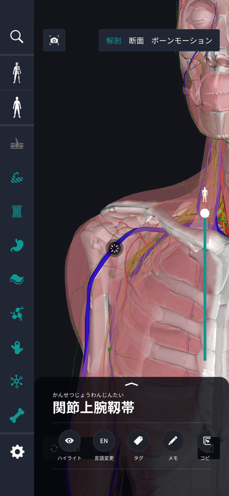
Textbook figures usually show the neck from the front, which hides how far back the greater horns reach. In the teamLabBody 3D anatomy app you can rotate and zoom the neck to see the U shape from above, and PREMIUM cross-sections show the hyoid, larynx and pharynx in a horizontal slice.
FAQ
- How can I feel my hyoid bone?
- Move a finger down from under your chin toward the Adam's apple. About one finger's width above it you will feel a hard bar. Pinch it from both sides and swallow: it moves up and down.
- Is the hyoid connected to any other bone?
- Not by a joint. The lesser horns hang from the styloid processes by the stylohyoid ligaments, and the body and greater horns are joined to the thyroid cartilage by the thyrohyoid membrane.
- Which muscles raise and lower the hyoid?
- The suprahyoid muscles (digastric, stylohyoid, mylohyoid, geniohyoid) raise it. The infrahyoid muscles (sternohyoid, omohyoid, sternothyroid, thyrohyoid) lower it; the sternothyroid attaches to the thyroid cartilage.