What are the abductors of the thumb (abductor pollicis longus and brevis)? Position, function and nerve supply
- Introduction
- What are the abductors of the thumb?
- How to read the name of the abductors of the thumb
- Features of the abductors of the thumb
- Where the abductors of the thumb are
- How to remember the abductors of the thumb
- The abductors of the thumb in English and Latin
- Trivia about the abductors of the thumb
- Structures related to the abductors of the thumb: features of the nerve supply
- Structures related to the abductors of the thumb: where the blood supply comes from
- Structures related to the abductors of the thumb: the anatomical snuffbox
- Quiz on the abductors of the thumb with answer
- Summary
Introduction
The abductors of the thumb are the muscles that spread the thumb away from the plane of the palm; in Japanese they are read boshi-gaitenkin. Under this name come the abductor pollicis longus, which arises from the back of the forearm, and the abductor pollicis brevis in the thenar eminence of the palm; they attach to the base of the first metacarpal and to the base of the proximal phalanx of the thumb respectively. This article explains their position, how to read the name, the English names, nerve supply and blood supply.
Use this article to deepen your understanding of the body.
Watch the video on the abductors of the thumb here (in Japanese)
Download teamLabBody Pro for free
A 3D anatomy app that shows every structure of the human body
Download teamLabBody Pro here!

What are the abductors of the thumb?
The abductors of the thumb are muscles essential to the dexterity and function of the thumb. Thanks to them we can carry out complex movements in daily life such as gripping objects and writing. They support the free movement of the thumb and greatly improve the function of the hand.
How to read the name of the abductors of the thumb
In Japanese the name is read boshi-gaitenkin and refers to the muscles that have the power to move the thumb (boshi) outward.
Features of the abductors of the thumb
The thumb is a very special part of the human hand: its bones differ from those of the other fingers and allow a wide variety of movements. The abductors of the thumb play an important role as the muscles that support the movement of spreading the thumb outward, that is, abduction.
Where the abductors of the thumb are
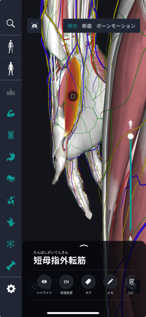
The abductor pollicis longus arises on the back of the forearm, from the posterior surfaces of the radius and ulna and the interosseous membrane, passes along the thumb side of the wrist and attaches to the base of the first metacarpal. The abductor pollicis brevis arises from the flexor retinaculum of the wrist and from the scaphoid and trapezium, and attaches to the lateral side of the base of the proximal phalanx of the thumb. Both contribute to abduction of the thumb and, by inserting at different sites, support the thumb's range of motion.
How to remember the abductors of the thumb
Combine "boshi = thumb" with "gaiten = movement outward": they are "the muscles that move the thumb outward". The key is to remember "long and short = from far and from near": the abductor pollicis longus is the long muscle coming from the forearm, the abductor pollicis brevis the short muscle within the palm.
The abductors of the thumb in English and Latin
In English they are written "Abductor Pollicis Longus" and "Abductor Pollicis Brevis"; Latin also uses "Longus" and "Brevis", meaning the "long" and "short" abductor of the thumb respectively.
Trivia about the abductors of the thumb
In human evolution, the ability to abduct the thumb was extremely important for using tools. This ability, which sets us apart from other animals, has influenced the development of human culture and technology.
Next we explain which nerves supply the abductors of the thumb and which vessels supply their blood. The abductor pollicis longus and the abductor pollicis brevis differ not only in position but also in nerve and blood vessel. Comparing the two side by side makes them easier to organise.
Structures related to the abductors of the thumb: features of the nerve supply
The abductor pollicis longus is supplied by the deep branch of the radial nerve (posterior interosseous nerve), like the other extensors on the back of the forearm. The abductor pollicis brevis, on the other hand, is supplied by the recurrent branch of the median nerve, together with the thenar muscles (the superficial head of the flexor pollicis brevis and the opponens pollicis). The fact that the two muscles that abduct the thumb are supplied by different nerves is often asked in examinations.
Structures related to the abductors of the thumb: where the blood supply comes from
The abductor pollicis longus receives blood from the posterior interosseous artery, which runs on the back of the forearm. The abductor pollicis brevis is supplied by the superficial palmar branch that the radial artery gives off at the wrist. The radial artery is the artery whose pulse you feel on the thumb side of the wrist; it then passes to the back of the hand, runs close to the base of the thumb and enters the deep part of the palm.
Structures related to the abductors of the thumb: the anatomical snuffbox
On the thumb side of the wrist, the tendon of the abductor pollicis longus, together with the tendon of the extensor pollicis brevis, forms the outer edge of the hollow that appears when you extend the thumb (the inner edge is the tendon of the extensor pollicis longus). This hollow is called the anatomical snuffbox, and at its floor lie the scaphoid and the radial artery.
The abductor pollicis brevis is the most superficial muscle of the thenar eminence; when you lift the thumb away from the plane of the palm you can feel it bulge. Display the abductor pollicis longus on the back of the forearm and the abductor pollicis brevis in the palm at the same time in 3D and see how the two muscles act on the same abduction of the thumb from different places.
Quiz on the abductors of the thumb with answer
Q: Which movement are the abductors of the thumb responsible for?
1. Bending the fingers
2. Straightening the fingers
3. Moving the thumb outward
4. Moving the thumb inward
A: 3. Moving the thumb outward
Correct answer: 3
As their name says, the abductors of the thumb make it possible to move the thumb outward.
Summary
This time we covered where the abductors of the thumb are, how to remember them and their names in English and Latin.
What did you think?
I hope this article has deepened your understanding of anatomy.
Learning is a long road with no end, but I sincerely wish you success. Let's keep learning together and preparing for the exam!
Look forward to the next article.
Learn more with the anatomy app teamLabBody Pro!
teamLabBody Pro is a 3D human anatomy app covering the whole body: muscles, organs, nerves, bones and joints.
The human body is faithfully reproduced from CT and MRI data of multiple subjects. Its medical-textbook-level content, supervised by doctors, can be viewed freely from any angle, so the app is useful in many medical settings, from explaining surgery to patients to anatomy study by students.
If you want to see the parts introduced here in more detail, download the anatomy app teamLabBody Pro.
Download teamLabBody Pro for free
A 3D anatomy app that shows every structure of the human body
Download teamLabBody Pro here!