beginning
In this article, I will explain the “kneecap” in detail. We'll delve into the kneecap and its associated tissues, particularly the important quadriceps femoris muscle. The quadriceps femoris muscle is an important muscle group whose four muscles contribute to knee extension and hip flexion, and is closely related to the kneecap. This article will focus on the characteristics, location, and relationship of the quadriceps femoris muscle to improve understanding of the structure and function of the knee joint. In particular, I will detail how the quadriceps femoris muscle and patella work together to support knee movement and contribute to physical movement. We'll also introduce some interesting trivia about the quadriceps muscle so that readers can gain knowledge to maintain a healthy knee joint.
Please use this article to deepen your understanding of the body.
Click here to watch a video about the kneecap
teamLab Body Pro Free Download
A 3D anatomy app that shows all the structures of the human body
Download teamLab Body Pro here!

What is the kneecap
The kneecap, or patella, is a small triangular bone in the knee joint, located in front of the knee, and plays a role in facilitating movement of the knee joint. It is located between the femur and tibia and plays an important role in helping stretch the knee in particular. Understanding the specific location and structure of the kneecap through human anatomy diagrams is very useful in learning the complex joint system of the human body.
How to read kneecap
The kneecap is read as “the key to discipline.”
Characteristics of the kneecap
The kneecap is characterized by its triangular shape and widening at the upper end. It is built into muscles, particularly the quadriceps femoris tendon, and is connected to the femur through the tendon. When the knee is bent, it functions like a pulley and plays a role in efficiently transferring muscle strength.
Location and position of the kneecap

The kneecap is located in front of the knee joint. If you look at the human anatomy chart, you can see that it is between the lower part of the femur and the upper part of the tibia, where it is sandwiched between the quadriceps femoris tendon and patellar tendon. This position contributes to the stability and motor ability of the knee joint.
How to remember a kneecap
It's a good idea to remember the kneecap as an image of a “knee shield.” It acts as a shield to mitigate impact from the front, and it is also an important part that plays a central role in moving the knee.
English and Latin for kneecap
The English notation for kneecap is “Patella,” and it is also expressed as “Patella” in Latin. These are expressions commonly used in the fields of medicine and anatomy.
Kneecap trivia
The kneecap is one of the hardest bones in the human body. As we get older, the surface of the kneecap loses its smoothness, which can be one of the causes of knee arthrosis. The size and shape of the kneecap varies from person to person, and this may affect knee pain.
Tissues associated with the kneecap: characteristics of the quadriceps femoris muscle
As the name suggests, the quadriceps femoris muscle consists of four muscles. These are called rectus femoris (rectus femoris), lateral broad muscle (Vastus Lateralis), medial broad muscle (Vastus intermedius), and medial medialis muscle (Vastus medialis). The main function of the quadriceps femoris muscle is to extend (extend) the knee, but when it comes to the straight hip muscle, it also participates in hip flexion (bending). The quadriceps femoris muscle has high endurance and strength, and enables movements such as walking, running, and jumping.
Tissue associated with the patella: location and position of quadriceps femoris muscle
The quadriceps femoris muscle is located at the front of the thigh (thigh). The straight crotch muscle passes through the front surface of the remaining three broad muscles and helps extend the leg forward. Most of the quadriceps femoris muscles are directly or indirectly related to the patella and transfer force to the patella through the quadriceps femoris tendon. The kneecap receives this force and plays an important role in helping the knee joint stretch.
Tissue associated with the kneecap: quadriceps trivia
The quadriceps is a very strong muscle group, but there is a risk of injury due to lack of exercise or improper training methods. In particular, pain around the knee is directly related to the quadriceps femoris muscle. The straight thigh muscle is the only quadriceps muscle that straddles the hip joint. This not only kicks the leg forward, but also plays a role in supporting posture when sitting. Vastus medialis muscle (Vastus medialis), particularly its raised part (VMO: Vastus Medialis Obliquus), is very important for knee stability and contributes to proper knee tracking movement.
A healthy relationship between the quadriceps femoris and patella is important for maintaining knee health and overall operational efficiency. Maintaining balance between these organizations through proper training and care is key to preventing injuries and improving performance.
Quiz about the kneecap
Q1: What is the main function of the kneecap?
— A: It helps stretch the knee and plays a role in facilitating movement of the knee joint.
Q2: Which part of the knee joint is the kneecap located?
— A: It is located in front of the knee joint.
summary
This time, I explained the location and location of the “kneecap”, how to memorize it, and the English/Latin notation.
How was it?
I would be happy if reading this article deepened my understanding of anatomy.
Learning is a long, never-ending journey, but I sincerely wish you all the best. Let's continue to study together and work hard for the national exam!
Please look forward to the next blog.
Learn more with the anatomy app “TeamLabBody Pro”!
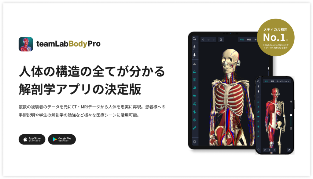
teamLabBody Pro is a “3D human anatomy application” that covers the entire body of the human body, including muscles, organs, nerves, bones and joints.
The human body is faithfully reproduced from CT and MRI data based on data from multiple subjects. Since medical book-level content supervised by physicians can be freely viewed from all angles, it can be used for various medical situations, such as explaining surgery to patients and studying anatomy for students.
If you want to see the parts introduced this time in more detail, please download the anatomy application “teamLabBody Pro.”
teamLab Body Pro Free Download
A 3D anatomy app that shows all the structures of the human body
Download teamLab Body Pro here!


