beginning
In this article, I will explain the “palmar aponeurosis” in detail. The palmar aponeurosis supports the structure of the palm of the hand and plays an important role in making the hand move smoothly. This membrane performs very important functions in daily life, such as reducing hand fatigue by helping hand movement and dispersing force. I will also touch on diseases and injuries that can occur in the palmar aponeurosis, and explain how to prevent them and treat them.
Please use this article to deepen your understanding of the body.
Click here to watch a video about palmar aponeurosis (palmar aponeurosis)
teamLab Body Pro Free Download
A 3D anatomy app that shows all the structures of the human body
Download teamLab Body Pro here!

What is palmar aponeurosis
The palmar aponeurosis is located in the palm of the hand and plays an important role in making the hand move smoothly and maintaining the shape of the hand. This aponeurosis is linked to multiple tendons, muscles, and bones, and is the foundation for realizing functional hand movement. In particular, it is important for acts such as grasping or pushing an object.
How to read palmar aponeurosis
Palmar aponeurosis is read as “shusho kenmaku” in Japanese. This comes from how to read the kanji, and it has the meaning of a membrane (aponeurosis) that wraps around tendons in the palm of the hand (palm).
Characteristics of palmar aponeurosis
The palmar aponeurosis is a very sturdy and elastic membrane. Therefore, palmar aponeurosis plays a role in protecting hands from various stresses that occur when using hands in daily life. It also reduces friction when bending and stretching fingers and supports smooth movement.
Location and location of palmar aponeurosis

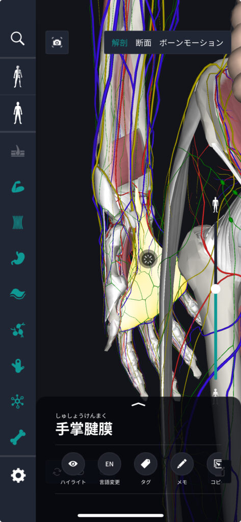
The palmar aponeurosis extends throughout the palm of the hand, particularly starting at the base of the fingers and extending deep into the palm. Each finger has its own aponeurosis, which cooperate with each other to support the movement of the palm of the hand.
How to remember palmar aponeurosis
In order to remember the location of the palmar aponeurosis, learning using a human anatomy diagram is effective. By expanding the palm of the hand and visualizing how aponeurosis spreads from the base of the finger to the deep part of the hand, it is possible to better understand its structure and function.
English and Latin for palmar aponeurosis
In English, palmar aponeurosis is expressed as “palmar fascia” or “palmar aponeurosis.” It is also often expressed as “aponeurosis palmaris” in Latin. If you know these words, it will be easier to gather information in international literature and materials.
Trivia about palmar aponeurosis
The palmar aponeurosis may become stiff due to aging or certain diseases. This can cause a condition called Dupuytren's contracture, where the fingers remain bent. Early treatment and prevention are important.
Tissues associated with palmar aponeurosis: characteristics of the carpal tunnel
The carpal tunnel (Carpal Tunnel) is a narrow channel at the base of the wrist. This passageway is surrounded by bone and carpal tunnel ligaments (thick ligaments that cross the wrist) and protect the median nerve and tendons that run inside. The palmar aponeurosis is particularly closely related to this carpal tunnel ligament and plays a role in supporting structures within the carpal tunnel. The median nerve, which passes through the carpal tunnel, controls sensory and muscular movements of the thumb, index finger, middle finger, and ring finger of the hand.
Tissues associated with palmar aponeurosis: location and location of carpal tunnel
The carpal tunnel is located on the palm side of the hand, at the bottom of the wrist. It is formed by small wrist bones called carpal bones and carpal tunnel ligaments that cover those bones. The palmar aponeurosis covers this area and indirectly affects the structure and function of the carpal tunnel. Tension or condition of the palmar aponeurosis can affect the space within the carpal tunnel and cause compression on the median nerve.
Tissues associated with palmar aponeurosis: carpal tunnel trivia
Carpal tunnel syndrome is caused by compression of the median nerve in a narrow passageway. Symptoms include numbness, pain, and impaired function of the fingers on the palm side of the hand. Hardening of the palmar aponeurosis increases pressure in the carpal tunnel and can be one of the causes of symptoms. Also, factors that increase the risk of carpal tunnel syndrome include repetitive hand movements, wrist inflammation, or excessive pressure on the wrist.
In order to prevent diseases related to palmar aponeurosis and carpal tunnel, it is effective to rest the wrist moderately and perform stretching and strengthening exercises. Also, improving the work environment can reduce the risk of carpal tunnel syndrome.
Through this article, I hope it will be an opportunity for you to deepen your understanding of the relationship between palmar aponeurosis and carpal tunnel and learn how to use your hands healthier.
Quiz about palmar aponeurosis
Q1: Where is the palmar aponeurosis located on the hand?
A. back of the hand
B. palm
C. Wrist
Correct answer: B. palm
Q2: What is the main function of the palmar aponeurosis?
A. Adjust the temperature of your hands
B. Improves grip
C. Protect your wrists
Correct answer: B. Improve your grip
summary
This time, I explained the location and location of the “palmar aponeurosis”, how to remember it, and the English and Latin notation.
How was it?
I would be happy if reading this article deepened my understanding of anatomy.
Learning is a long, never-ending journey, but I sincerely wish you all the best. Let's continue to study together and work hard for the national exam!
Please look forward to the next blog.
Learn more with the anatomy app “TeamLabBody Pro”!
teamLabBody Pro is a “3D human anatomy application” that covers the entire body of the human body, including muscles, organs, nerves, bones and joints.
The human body is faithfully reproduced from CT and MRI data based on data from multiple subjects. Since medical book-level content supervised by physicians can be freely viewed from all angles, it can be used for various medical situations, such as explaining surgery to patients and studying anatomy for students.
If you want to see the parts introduced this time in more detail, please download the anatomy application “teamLabBody Pro.”
teamLab Body Pro Free Download
A 3D anatomy app that shows all the structures of the human body
Download teamLab Body Pro here!


