beginning
In this article, I will explain “rotator cuffs” in detail.
The rotator cuff is an important structure that controls the stability and motor ability of the shoulder joint, and is formed by combining the tendons of multiple muscles. This article explains a wide range of rotator cuff anatomy, causes and symptoms of rotator cuff injury, and its prevention and treatment. In particular, rotator cuff injuries seen in athletes and the elderly can have a major impact on daily life, but they can be managed with proper knowledge and care. Please use this article to deepen your understanding of the body.
Click here to watch a video about rotator cuffs (kemban)
teamLab Body Pro Free Download
A 3D anatomy app that shows all the structures of the human body
Download teamLab Body Pro here!

What is a rotator cuff
A rotator cuff is a collection of muscle tendons that play an important role in supporting stability and mobility around the shoulder joint. These tendons are arranged to cover the head of the humerus bone constituting the shoulder, making the movement of the shoulder joint smooth and preventing dislocations during strong movements. In an explanation using a human anatomy diagram, you can clearly understand how important this rotator cuff is for shoulder joint function.
How to read rotator cuff
The rotator cuff is read as “good fight” in Japanese. This way of reading is a combination of a tissue called a tendon (tendon) and a shape called a plate (band), and it literally comes from the fact that tendons are structured like plates.
Characteristics of rotator cuffs
The biggest characteristic of a rotator cuff is that the four main tendons that are its components, namely the supraspinatus tendon, subspinous tendon, small circular tendon, and subscapula tendon, work closely together to support the movement of the shoulder joint. These tendons work uniquely while being close to each other, making complex shoulder movements possible.
Location and position of the rotator cuff

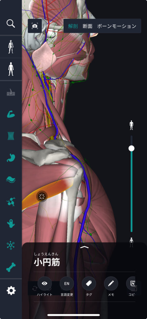
The rotator cuff is arranged to surround the humerus head of the shoulder joint. If you look at the human anatomy chart, you can see how the rotator cuff covers the spherical head of the humerus bone, and you can visually confirm how it contributes to stabilizing the shoulder joint.
(The rotator cuff is composed of the four main tendons, “supraspinatus tendon,” “subspinous tendon,” “small circular tendon,” and “subscapular tendon,” and the “small circular tendon” is displayed.)
How to remember a rotator cuff
As a way to remember the four tendons that make up a rotator cuff, there is a method of taking the acronym for each muscle and memorizing it as “spinous shoulder.” This is a combination of the initials supraspinatus muscle (supraspinatus), subspinous muscle (s), small circle muscle (s), and subscapula muscle (scapula muscle).
English/Latin for rotator cuff
The rotator cuff is expressed as “Rotator Cuff” in English. It is called “Manschette rotatoria” in Latin, and both indicate a collection of tendons that support the function of the shoulder joint called the rotator tendon ring.
Trivia about rotator cuffs
Rotator cuff injuries are a common problem in athletes and older people who overuse their shoulders. Early detection and proper treatment are necessary, and proper shoulder joint preservation measures and strength training are recommended for prevention.
Tissues associated with rotator cuffs: rotator cuff characteristics
The rotator cuff is mainly composed of 4 muscles: supraspinatus (Supraspinatus), infraspinatus (Infraspinatus), teres minor (Teres Minor), and subscapularis (Subscapularis). All of these are attached to the humerus by tendons, which support the smooth movement of the shoulder joint and prevent shoulder dislocation and friction damage.
Each rotator cuff muscle performs a specific function by moving the shoulder joint in different directions. For example, the supraspinatus muscle helps lift the arm, the infraspinatus muscle and the circumflex minor muscle support external rotation of the shoulder, and the subscapula muscle is responsible for internal rotation.
Tissues related to rotator cuffs: rotator cuff location/position
The rotator cuff is located deep in the shoulder joint, specifically between the head of the humerus bone and the shoulder blade. This arrangement makes it possible to perform movements such as raising the arm, rotating it, and pulling it to the body in a stable manner. If you look at the human anatomy diagram, you can clearly see how these muscles are arranged around the shoulder joint. Actually, the arrangement of the muscles is very reasonable and provides maximum range of motion and stability for the shoulder.
Tissues associated with rotator cuffs: rotator cuff trivia
The rotator cuff is one of the areas with a high risk of injury, especially in athletes and people who perform manual labor. The act of frequently lifting the arm overhead puts stress on the supraspinatus muscle in particular and can cause inflammation or rupture. Also, with age, the muscles and tendons of the rotator cuff weaken more easily, which can cause pain and dysfunction. However, it is possible to minimize these risks by using proper strength training and techniques to protect the shoulder joint.
Rotator cuff quiz and correct answers
Q1.What are the four tendons that make up the rotator cuff?
Correct answer: These are supraspinatus tendons, subspinous tendons, small round tendons, and subscapular tendons.
summary
This time, I explained the location and position of the “rotator cuff”, how to memorize it, and the English and Latin notation.
How was it?
I would be happy if reading this article deepened my understanding of anatomy.
Learning is a long, never-ending journey, but I sincerely wish you all the best. Let's continue to study together and work hard for the national exam!
Please look forward to the next blog.
Learn more with the anatomy app “TeamLabBody Pro”!
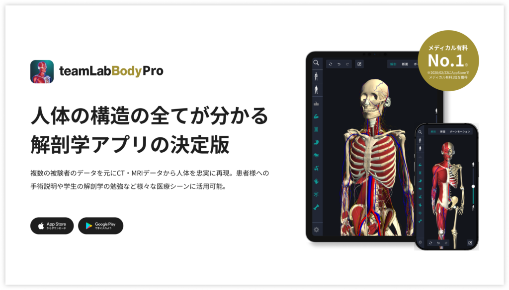
teamLabBody Pro is a “3D human anatomy application” that covers the entire body of the human body, including muscles, organs, nerves, bones and joints.
The human body is faithfully reproduced from CT and MRI data based on data from multiple subjects. Since medical book-level content supervised by physicians can be freely viewed from all angles, it can be used for various medical situations, such as explaining surgery to patients and studying anatomy for students.
If you want to see the parts introduced this time in more detail, please download the anatomy application “teamLabBody Pro.”
teamLab Body Pro Free Download
A 3D anatomy app that shows all the structures of the human body
Download teamLab Body Pro here!


