beginning
In this article, I will explain the “trapezius muscle” in detail. The trapezius muscle extends to the neck, shoulders, and back and plays an important role in supporting movement and posture. This muscle, which is read as “sobokin,” is similar in shape to an old monk's hat, and has characteristics in size and the three parts of the upper, middle, and lower parts. Also, it is called “Trapezius muscle” in English and “Musculus trapezius” in Latin, and its shape is reminiscent of a “trapezoid.” Please use this article to deepen your understanding of the body.
Click here to watch a video about the trapezius muscle (trapezius muscle)
teamLab Body Pro Free Download
A 3D anatomy app that shows all the structures of the human body
Download teamLab Body Pro here!

What is the trapezius muscle
The trapezius muscle is a large muscle that extends to the neck, shoulders, and back, and has an important role in supporting movement and posture. The trapezius muscle is essential for stabilizing movement, especially around the neck and shoulders. The name of the trapezius muscle comes from its shape similar to the hat worn by monks in the old days, or “trapezius.”
How to read trapezius
The trapezius muscle is read as “sobokin.”
Characteristics of the trapezius muscle
The characteristics of the trapezius muscle are its size and the fact that it has a triangular shape and is divided into 3 parts: upper part, middle part, and lower part. Each part has muscle fibers in different directions, making it possible to support various movements.
Location and position of the trapezius muscle

According to the human anatomy diagram, the trapezius muscle starts at the back of the head, that is, at the bottom of the neck, and extends above the shoulder blades. The middle part extends sideways along the upper edge of the shoulder blades, and the lower part extends from the inside of the shoulder blades through the center of the back and close to the lower back. Due to their widespread arrangement, the trapezius muscle affects multiple movements of the neck, shoulders, and back.
How to remember the trapezius muscle
Remember that it's named because its shape resembles a trapezoid. Another important point is that it is divided into 3 parts from top to bottom.
English and Latin for trapezius
The “trapezius muscle” is called “trapezius muscle” in English and “musculus trapezius” in Latin. The name comes from the fact that the shape of the muscle is reminiscent of a geometric shape, particularly a “trapezoid.”
Trapezius muscle trivia
Here's some trivia.
The trapezius muscle is one of the muscles that is susceptible to stress, and improper posture due to prolonged desk work or smartphone use can cause excessive muscle tension. This may cause stiff shoulders and neck pain. It is important to reduce muscle tension by doing regular stretches and massages.
Tissues associated with the trapezius muscle: characteristics of the cervical spine
The cervical spine is part of the spine and plays a role in supporting the head. The cervical spine connects the upper skull to the lower thoracic spine. A person's cervical spine usually consists of 7 vertebrae, and each vertebra is numbered from C1 to C7. Compared to other spine parts, the cervical spine can move more finely, increasing the range of motion of the neck.
In particular, the relationship between the cervical spine and the trapezius muscle is deep, and the trapezius muscle is a large muscle that starts in the cervical spine and reaches the shoulder blades. The trapezius muscle supports neck and shoulder movements and plays a role in maintaining head posture, and if the cervical spine is not healthy, the trapezius muscle may also be affected.
Tissues related to the trapezius muscle: location and position of the cervical spine
The cervical spine is located from the lower side of the head to the upper part of the chest. The uppermost vertebra (C1) is called the “atlas” and has a disc-shaped structure that supports the skull. The next vertebra (C2) is called an “axis,” and in combination with an atlas, it has the function of rotating the head left and right. Such a special structure allows the head to move in multiple directions.
Tissues associated with the trapezius muscle: trivia about the cervical spine
The cervical spine is an area prone to injury due to its wide range of motion. Whiplash, which occurs when you are involved in a traffic accident, occurs as a result of a sudden sharp movement of the neck bone back and forth. Also, if you continue to maintain an unnatural posture by working at a desk or using a smartphone for a long time, the load on your neck bones will increase. Therefore, it is important to keep your neck bones and trapezius muscles, or back muscles, healthy by stretching frequently and exercising moderately.
The neck bones and the trapezius muscle are closely related. For example, neck bone problems may cause pain or tension in the trapezius muscles, and conversely, when the trapezius muscle is excessively tense, it can put a strain on the neck bone and cause symptoms such as a herniated disc. In order to prevent neck bone and trapezius muscle problems, it is effective to keep a correct posture on a regular basis and relax your muscles regularly.
Trapezius Quiz
Q: What part of the body is the trapezius muscle located in?
A: neck, shoulders, back
summary
This time, I explained the location and location of the “trapezius muscle”, how to memorize it, and the English and Latin notation.
How was it?
I would be happy if reading this article deepened my understanding of anatomy.
Learning is a long, never-ending journey, but I sincerely wish you all the best. Let's continue to study together and work hard for the national exam!
Please look forward to the next blog.
Learn more with the anatomy app “TeamLabBody Pro”!
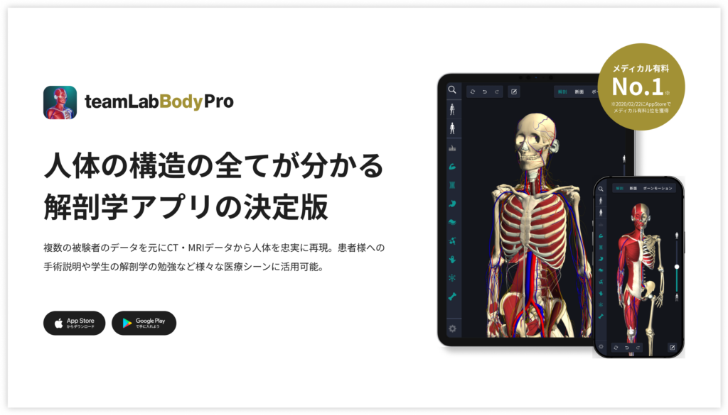
teamLabBody Pro is a “3D human anatomy application” that covers the entire body of the human body, including muscles, organs, nerves, bones and joints.
The human body is faithfully reproduced from CT and MRI data based on data from multiple subjects. Since medical book-level content supervised by physicians can be freely viewed from all angles, it can be used for various medical situations, such as explaining surgery to patients and studying anatomy for students.
If you want to see the parts introduced this time in more detail, please download the anatomy application “teamLabBody Pro.”
teamLab Body Pro Free Download
A 3D anatomy app that shows all the structures of the human body
Download teamLab Body Pro here!


