
Cartilage: where it is found, its types and the nerves around it
Introduction
In this article we explain effective ways to study human anatomy.
In human anatomy you have to remember not only the names of the organs, muscles and bones, but also where in the body they are. That is why it pays to study as efficiently as possible.
This time we explain how to study cartilage.
Download teamLabBody Pro for free
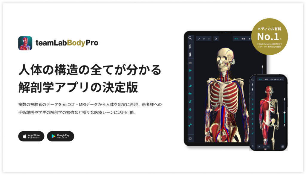
A 3D anatomy app that shows every structure of the human body
Download teamLabBody Pro here!
Learning with an anatomy app
In the anatomy app you can view a curated set of 3D anatomical models. Each model can be observed in several ways: surface, cross sections, the nervous system and more.
1. Where cartilage is found

Cartilage is found widely throughout the body. The main places are the ends of the bones in joints, the front ends of the ribs (where they join the sternum), the outer ear, the tip of the nose, and the larynx and trachea.
In joints, cartilage covers the ends of the bones, reduces friction and allows smooth movement. This is called articular cartilage.
The cartilage of the ribs connects the ribs to the sternum and helps the rib cage move during breathing. The cartilage of the ear and the nose is known as auricular cartilage and nasal cartilage, and it helps these parts keep their shape and flexibility. The cartilages of the larynx assist the movement of the vocal folds and support the production of voice.
The cartilage rings of the trachea keep the airway open and are important for smooth breathing. According to the type of fibres, cartilage is divided into three kinds: hyaline cartilage (articular, costal and tracheal cartilage), elastic cartilage (the auricle and the epiglottis) and fibrocartilage (the intervertebral discs and the pubic symphysis).
2. Muscles related to cartilage

Cartilage itself contains no muscle, but there are muscles around it that support movement. Around the articular cartilage of the knee and the elbow, for example, are the quadriceps femoris and the biceps brachii, which move these joints.
As these muscles contract and relax, they help make movement smooth while friction is kept low.
The muscles that work together with the cartilage around the ribs include the diaphragm and the intercostal muscles, which support breathing.
In the ear and nose there are few muscles attached specifically to these cartilages, but the facial muscles of expression support their shape and movement. This supports the small movements of the nose and ears during facial expression and speech.
3. The main nerves around cartilage

Cartilage itself has neither blood vessels nor nerves. Nutrients reach it by diffusion: in articular cartilage from the synovial fluid, and in other cartilage from the vessels of the perichondrium that covers its surface. So knowing the nerves of the tissues around the cartilage is the key to understanding the sensations related to it.
Around articular cartilage, sensory nerves are distributed to the joint capsule, the ligaments and the subchondral bone beneath the cartilage, and they sense the position of the joint and the pressure on it. At the knee, articular branches of the femoral, obturator and tibial nerves reach the joint capsule; at the elbow, articular branches of the musculocutaneous, median, radial and ulnar nerves.
Sensation from the skin covering the auricular cartilage is carried by the great auricular nerve of the cervical plexus and the auriculotemporal nerve of the trigeminal nerve. The skin covering the nasal cartilages and the mucosa of the nasal cavity are supplied by branches of the ophthalmic and maxillary nerves, both divisions of the trigeminal nerve.
Around the cartilages of the larynx, the internal branch of the superior laryngeal nerve carries sensation from the mucosa above the glottis, and the recurrent laryngeal nerve carries sensation from the mucosa below the glottis and controls the muscles of the larynx. Display the cartilage in 3D and overlay the nerves around it to see the relationship: "cartilage has no nerves; the surrounding tissues provide the sensation".
Concrete ways to study with the app
Here are concrete ways to study using a human anatomy app.
Review your learning history and practice repeatedly
These are the steps for reviewing your anatomy learning history and revising effectively.
1. Check your learning history in the app
Checking your study history in the app is an important step for making effective progress in anatomy. First open the app and go to the history section from the main menu. Many anatomy apps show your progress in graphs or lists, so you can see which parts you have studied and how much time you spent on each.
With that data you can see where you are strong and where you need to invest more time and effort. It also helps to mark the parts you find difficult or need to relearn with tags or notes. Checking your history regularly and going back over what you have already studied leads to effective review and deeper understanding.
2. Plan your revision
Planning efficient revision from your learning history is extremely effective for making knowledge stick. First identify your weak points and the topics you need to relearn. Then spread these items across a weekly or monthly calendar and create a concrete study schedule. Working to a plan lets you cover every region evenly and avoids cramming a large amount of information at once.
It helps to use a task management app or a digital calendar to set study reminders. It is also important to review your progress regularly and stay flexible enough to adjust the plan when needed. Studying with a goal and a plan is the efficient way to acquire anatomical knowledge.
3. Learn visually with the 3D functions
The 3D functions make anatomy much easier to understand visually. A 3D model shows the structure of the body in three dimensions and lets you observe each part in detail. This gives you an intuitive grasp of the positional relationships of deep muscles and organs that are hard to see in a flat diagram. For example, you can rotate a particular muscle or bone and zoom in and out to study it down to the smallest detail.
Many apps can also display cross sections of each region on the 3D model, which helps you understand internal structure. This variety of visual information aids memory and improves your ability to respond quickly in exams and in practice. Learning visually with 3D lets you acquire anatomical knowledge more deeply and more efficiently.
Make good use of the memo function

Make notes of what you notice and the key points while you study so that you do not forget them. The memo function offers text input, saved images and handwritten notes, so you can use whichever suits your purpose. Tag your memos so that they are easy to find later.
Test yourself regularly with quizzes
Testing what you have learned regularly in quiz form is a very effective way to make anatomical knowledge stick. Quizzes let you repeat the material while giving you an objective view of how well you understand it and where the gaps are.
For example, taking quizzes in a study app at set intervals lets you recheck what you have learned and strengthen your memory. Quiz formats range from multiple choice to fill-in-the-blank and short answer, and each one helps you understand from a different angle and draw on different kinds of knowledge.
Get feedback
If you can, ask other learners or specialists for feedback. It helps you find what you have misunderstood and what you can improve. Testing yourself regularly also keeps your motivation up: a sense of achievement and progress makes you want to keep learning.
Summary
This time we explained how to study "cartilage" with an app!
Thank you for reading this far.
I hope this article helps you with your study of anatomy.
Learning is a long road with no end, but I sincerely wish you success. Let's keep learning together and work toward the exam!
Look forward to the next article.
Download teamLabBody Pro for free
A 3D anatomy app that shows every structure of the human body
Download teamLabBody Pro here!


