
Striated muscle: where it is, its structure and its nerves
Introduction
Here is how to study human anatomy efficiently.
In human anatomy you have to remember not only the names of the organs, muscles and bones, but also where in the body they are. That is why it pays to study as efficiently as possible.
This time, we explain how to study striated muscle.
Download teamLabBody Pro for free
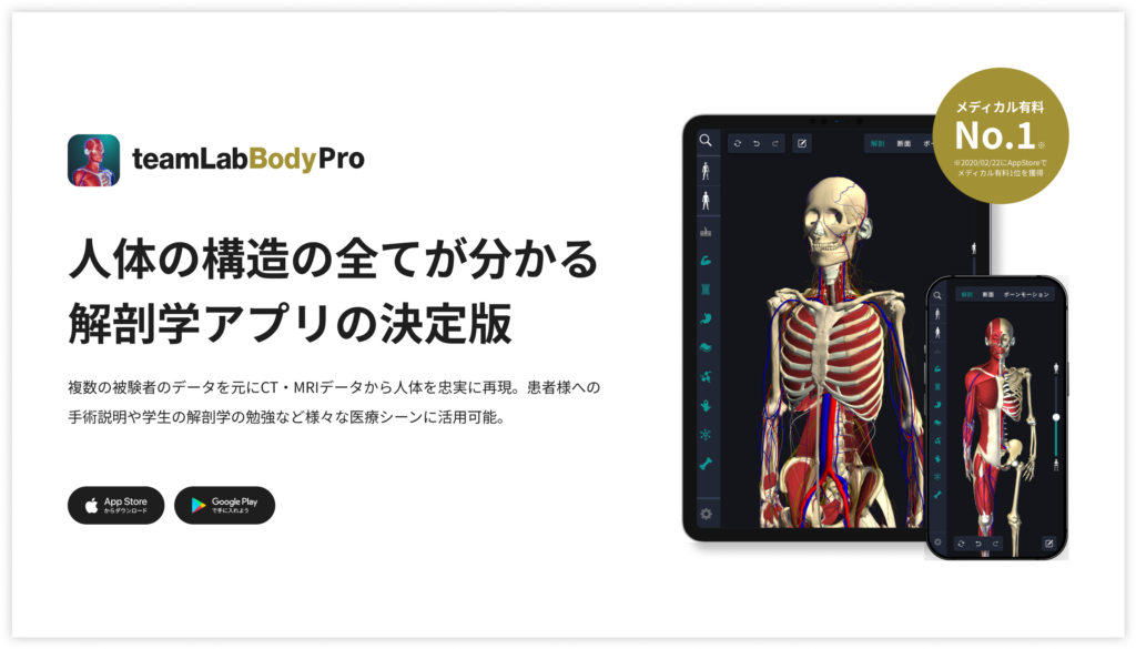
A 3D anatomy app that shows every structure of the human body
Download teamLabBody Pro here!
Learning with an anatomy app
In the anatomy app you can view a curated set of 3D anatomical models. Each model can be observed in several ways: surface, cross sections, the nervous system and more.
1. Where striated muscle is found

Striated muscle includes skeletal muscle and cardiac muscle. Of these, skeletal muscle is found widely throughout the body; it is mostly attached to bone and can be controlled voluntarily. These muscles play an important role in movement and in changing body position.
Specifically, skeletal muscle is found in the limbs (such as the quadriceps femoris and biceps brachii), in the trunk (such as the rectus abdominis and trapezius) and in the face (such as the buccinator among the muscles of facial expression and the masseter among the muscles of mastication).
The main function of skeletal muscle is to move bones by moving joints, which makes movement possible. It also plays an important role in maintaining posture. In addition, skeletal muscle produces heat and contributes to regulating body temperature.
Understanding where each striated muscle is located is basic knowledge in anatomy and is very important in medicine, physical therapy and sports science.
2. The components of striated muscle

Striated muscle is made up of various components, including muscle fibers, connective tissue, blood vessels and nerves. Muscle fibers are long, thin cells that are divided into even finer structures called myofibrils.
Myofibrils are made of sarcomeres composed of the proteins actin and myosin, and these form the striped pattern characteristic of striated muscle.
Muscle contraction is produced by the sliding movement within these sarcomeres. Connective tissue bundles the muscle fibers, supports the muscle as a whole and absorbs external shocks.
Blood vessels supply the muscle with oxygen and nutrients and remove waste products. Nerves transmit the commands for contraction and make precise control of movement possible. When these components work in harmony, striated muscle functions efficiently and can carry out complex movements.
3. The main nerves of striated muscle

The main nerves of striated muscle are motor nerves, many of which come from the spinal nerves. Spinal nerves are distributed to every part of the body and control the specific functions of each muscle.
For example, the biceps brachii is supplied by the musculocutaneous nerve, and the femoral nerve plays the main role for the quadriceps femoris. These motor nerves carry signals from the brain to the muscles and make them contract.
Sensory nerves are also involved with striated muscle, reporting the state of the muscle to the brain. This two-way exchange of information makes precise control of movement possible.
In addition, the diameter of the blood vessels inside the muscle is adjusted by the sympathetic nerves, so blood flow changes to match activity (skeletal muscle receives no parasympathetic supply). This complex network of the nervous system supports the advanced motor function of striated muscle.
Concrete ways to study with the app
Here are concrete ways to study using a human anatomy app.
Review your learning history and practice repeatedly
These are the steps for reviewing your anatomy learning history and revising effectively.
1. Check your learning history in the app
Checking your study history in the app is an important step for making effective progress in anatomy. First open the app and go to the history section from the main menu. Many anatomy apps show your progress in graphs or lists, so you can see which parts you have studied and how much time you spent on each.
With that data you can see where you are strong and where you need to invest more time and effort. It also helps to mark the parts you find difficult or need to relearn with tags or notes. Checking your history regularly and going back over what you have already studied leads to effective review and deeper understanding.
2. Plan your revision
Planning efficient revision from your learning history is extremely effective for making knowledge stick. First identify your weak points and the topics you need to relearn. Then spread these items across a weekly or monthly calendar and create a concrete study schedule. Working to a plan lets you cover every region evenly and avoids cramming a large amount of information at once.
It helps to use a task management app or a digital calendar to set study reminders. It is also important to review your progress regularly and stay flexible enough to adjust the plan when needed. Studying with a goal and a plan is the efficient way to acquire anatomical knowledge.
3. Learn visually with the 3D functions
The 3D functions make anatomy much easier to understand visually. A 3D model shows the structure of the body in three dimensions and lets you observe each part in detail. This gives you an intuitive grasp of the positional relationships of deep muscles and organs that are hard to see in a flat diagram. For example, you can rotate a particular muscle or bone and zoom in and out to study it down to the smallest detail.
Many apps can also display cross sections of each region on the 3D model, which helps you understand internal structure. This variety of visual information aids memory and improves your ability to respond quickly in exams and in practice. Learning visually with 3D lets you acquire anatomical knowledge more deeply and more efficiently.
Make good use of the memo function

Make notes of what you notice and the key points while you study so that you do not forget them. The memo function offers text input, saved images and handwritten notes, so you can use whichever suits your purpose. Tag your memos so that they are easy to find later.
Test yourself regularly with quizzes
Testing what you have learned regularly in quiz form is a very effective way to make anatomical knowledge stick. Quizzes let you repeat the material while giving you an objective view of how well you understand it and where the gaps are.
For example, taking quizzes in a study app at set intervals lets you recheck what you have learned and strengthen your memory. Quiz formats range from multiple choice to fill-in-the-blank and short answer, and each one helps you understand from a different angle and draw on different kinds of knowledge.
Get feedback
If you can, ask other learners or specialists for feedback. It helps you find what you have misunderstood and what you can improve. Testing yourself regularly also keeps your motivation up: a sense of achievement and progress makes you want to keep learning.
Summary
This time we looked at how to study striated muscle using an app!
Thank you for reading this far.
I hope this article helps you with your study of anatomy.
Learning is a long road with no end, but I sincerely wish you success. Let's keep learning together and work toward the exam!
Look forward to the next article.
Download teamLabBody Pro for free
A 3D anatomy app that shows every structure of the human body
Download teamLabBody Pro here!


