beginning
In this article, I will explain the “Achilles heel” in detail. The Achilles tendon is a strong, thick tendon that connects to the heel bone and transmits muscle strength around the ankle. The Achilles tendon is involved in foot movements such as walking, running, and jumping, and the heel bone is the largest and strongest bone in the human foot. This article explains how to read the Achilles tendon, its location, characteristics, English and Latin expressions, and the related tissue calcaneus. It also introduces trivia and quizzes about the Achilles tendon. Please use this article to deepen your understanding of the body.
Click here to watch a video about Achilles tendon (Achilles tendon)
teamLab Body Pro Free Download
A 3D anatomy app that shows all the structures of the human body
Download teamLab Body Pro here!

What is the Achilles tendon
The Achilles tendon is the strongest and thickest tendon in the human lower leg. This tendon connects to the heel bone (heel bone) and transfers strength to the muscles around the ankle (especially the calf muscles, that is, gastrocnemius and soleus muscles). The Achilles tendon is an important tendon involved in foot movements, such as walking, running, and jumping.
How to read Achilles heel
“Achilles heel” is read as “Achilles' heel.” “Achilles” comes from the Greek mythology hero Achilles.
Characteristics of the Achilles tendon
The Achilles tendon is a very strong and thick tendon that transmits muscle strength around the ankle and is involved in foot movements such as walking, running, and jumping.
The location and position of the Achilles tendon

The Achilles tendon is behind the foot, just above the ankle. It is particularly characteristic that it is fixed to the protruding part at the top of the heel bone (heel bone). By looking at an anatomical diagram of the human body, it is possible to specifically understand the location and shape of the Achilles tendon.
How to remember the Achilles tendon
It's good to remember that the Achilles heel is named after the Greek mythological hero Achilles. Achilles held his heel when his mother Thetis immersed Achilles in the River Styx in order to immortalize him, so only the heel part was not soaked, and that was regarded as his weak point. It is named after this legend.
English and Latin for Achilles tendon
The Achilles tendon is called Achilles tendon in English. Also, in Latin, it is expressed as Tendo calcaneus. Tendo means tendon, and calcaneus means calcaneus.
Achilles tendon trivia
Here's some trivia.
The Achilles tendon is a very strong structure, but it can be damaged or torn if too much stress is applied during exercise. This is more likely to happen in athletes and active people and requires proper treatment and rehabilitation. Also, an Achilles tendon rupture is generally regarded as an injury common among men in their 30s to 40s.
Tissues associated with the Achilles tendon: characteristics of the calcaneus
The heel bone (heel bone) is the largest and strongest bone in the human foot, and is located on the back side of the foot. This bone plays a very important role in successfully accepting the force applied to the foot when walking or running. Thanks to the heel bone, strength can be transmitted firmly to the ankle via the Achilles tendon. Also, since the heel bone is strong and absorbs shock well, it reduces pain and discomfort when walking or running.
Tissues associated with the Achilles tendon: location and position of the heel bone
The calcaneus is located at the back of the foot bones. This bone plays a role in supporting our body weight and stabilizing our ankles. Furthermore, the calcaneus is connected to the Achilles tendon and works together with the gastrocunemius muscle and soleus muscle, which are calf muscles, to help with the movement of bending the ankle.
Tissues associated with the Achilles tendon: trivia about the calcaneus
Here's some trivia.
1. The heel bone is the largest of the 26 bones in the foot. Also, since the shape differs from person to person, this is an important point when comparing foot types.
2. The calcaneus plays an important role in paleontological and anthropological research. By investigating the shape of the calcaneus from biological footprints and fossils, it is possible to estimate how living things walk and how they evolve.
3. One cause of heel bone pain is a bone spur called a heel spur (heel spur). This is a symptom of abnormal bone spines occurring on the surface of the heel bone, and may cause a condition similar to pain due to Achilles tendonitis or plantar aponeurosis.
4. The heel bone can be damaged by exercise or injury. A heel fracture occurs when an impact is applied to the ankle, and treatment is performed with rest, braces, or surgery.
In this way, the heel bone, which is closely related to the Achilles tendon, plays an important role in foot function and stability, and can be said to be an essential tissue when we walk and exercise. In order to keep your feet healthy, it is essential to take care of your heel bone and Achilles tendon, so be aware of it in your daily life.
Achilles tendon quiz
Q: What is the origin of the name Achilles tendon?
A: It is named after the Greek mythological hero Achilles.
Q: What bone is the Achilles tendon connected to?
A: It is connected to the calcaneus (heel bone).
Q: What are the main muscles the Achilles tendon connects to?
A: These are gastrocnemius muscle and soleus muscle.
summary
This time, I explained the location and location of the “Achilles heel”, how to memorize it, and the English and Latin notation.
How was it?
I would be happy if reading this article deepened my understanding of anatomy.
Learning is a long, never-ending journey, but I sincerely wish you all the best. Let's continue to study together and work hard for the national exam!
Please look forward to the next blog.
Learn more with the anatomy app “TeamLabBody Pro”!
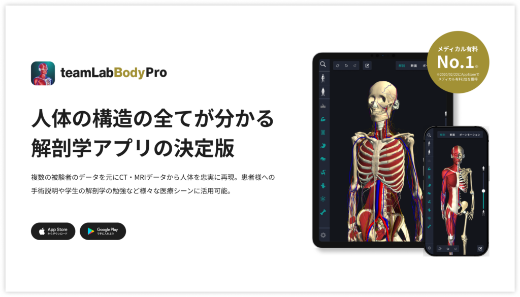
teamLabBody Pro is a “3D human anatomy application” that covers the entire body of the human body, including muscles, organs, nerves, bones and joints.
The human body is faithfully reproduced from CT and MRI data based on data from multiple subjects. Since medical book-level content supervised by physicians can be freely viewed from all angles, it can be used for various medical situations, such as explaining surgery to patients and studying anatomy for students.
If you want to see the parts introduced this time in more detail, please download the anatomy application “teamLabBody Pro.”
■teamLab Body Pro Free Download


