beginning
In this article, I will explain effective study methods, starting with knowledge of specialized parts in human anatomy.
In human anatomy, it is necessary not only to memorize the names of various organs, muscles, and bones, but also to remember where they are located in the body. Therefore, it is necessary to learn as efficiently as possible.
I hope you can read this article and use the app to deepen your understanding even a little bit.
Now, I'll explain the details about the “optic nerve” and how to study human anatomy.
teamLab Body Pro Free Download
A 3D anatomy app that shows all the structures of the human body
Download teamLab Body Pro here!

What is the optic nerve?
In the anatomy application, you can view a selection of anatomy 3D models. In this model, there are various observation methods such as surfaces, cross-sections, and nervous systems. This time, I'll explain using an anatomy application.
About the optic nerve

Study points
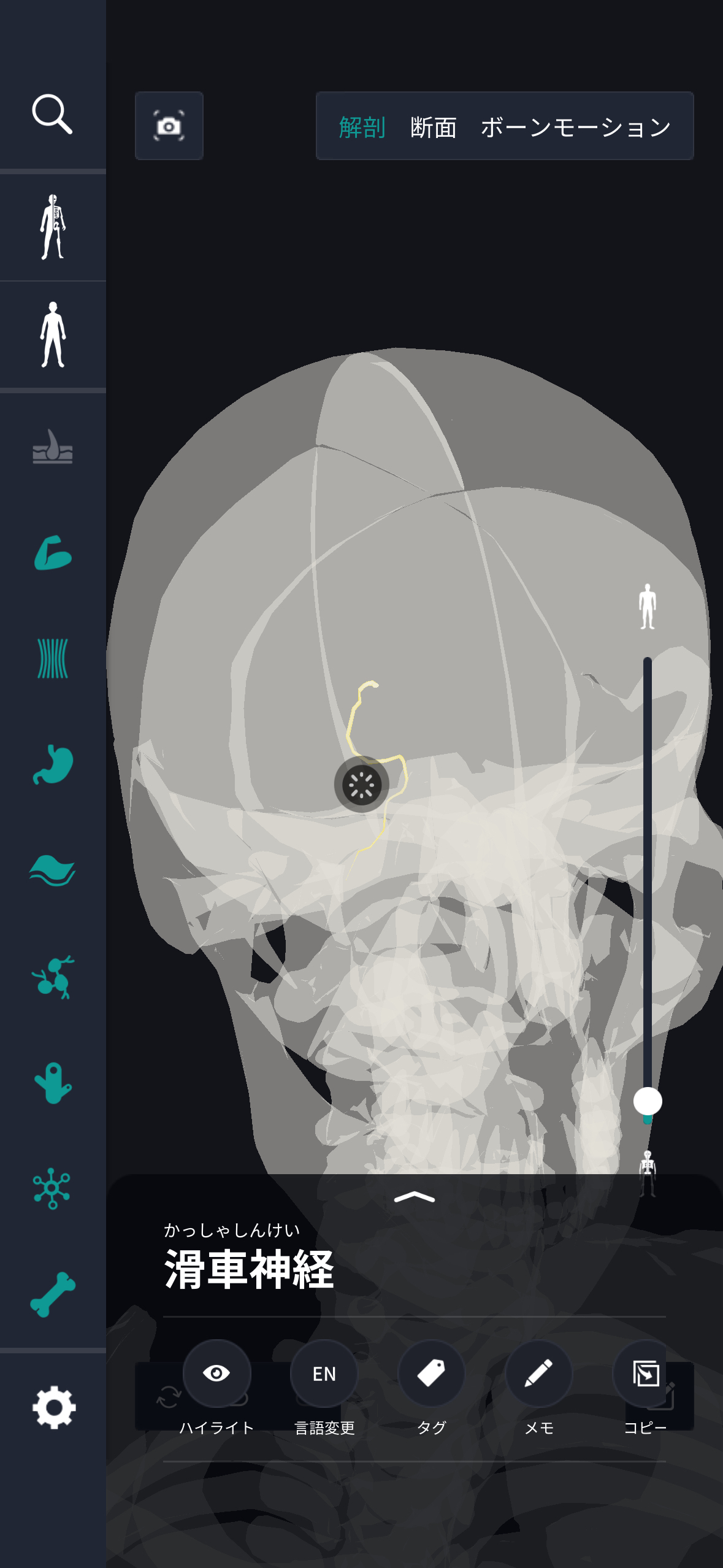
Location and structure of the optic nerve
The location and structure of the trochlear nerve is fascinating from an anatomical point of view. This nerve is the only cranial nerve that develops from the dorsal side of the brain stem, and is particularly unique in that it appears from the dorsal side of the bridge (pons) at the bottom of the midbrain. When exiting the brain stem, the trochlear nerve orbits the posterior brain cover, then runs forward along the dura mater, and passes through the superior orbital fissure to enter the orbital cavity. This complex pathway shows how nerves cleverly pass between various brain structures, and is part of the process leading to specific muscles. The trochlear nerve is characterized by its length and thinness, and connects specifically to the superior oblique muscle of the eyeball. As a structure, it is mainly composed of nerve fibers that transmit action potentials, and these fibers are not covered with myelin, and can quickly transmit action potentials. The trochlear nerve functions as a nerve that plays a specific role due to its arrangement and movement. Anatomically, these very fine structures and pathways of nerves can be viewed as advanced adaptations of biological design. Its precise position and structure are extremely important for controlling and coordinating eye movements, and these properties play a role in the trochlear nerve.
The role and function of the optic nerve
Looking at the role and function of the trochlear nerve from an anatomical perspective, there are several important factors to consider. This nerve is mainly developed to carry nerve signals that move the superior oblique muscle of the eyeball. The superior oblique muscle moves the eyeball downward and outward to coordinate vertical movement in an inverted state. Thus, the trochlear nerve plays an important role in fine-tuning eye movements. This movement contributes to the stability of the line of sight and accurate recognition of objects when looking at objects, and is essential when coordinating eye movements. Trochlear nerves exist on both sides of the body, and are particularly characteristic that they intersect and control the opposite eye. As a result, vision is controlled by the dual nervous system, and it has been confirmed that the movements of both eyes are effectively adjusted. Also, since this nerve can quickly process input related to movement, smooth visual tracking is possible. Therefore, the normal function of the trochlear nerve is part of visual search and spatial cognition, and it has a very useful function in everyday life. The role and function of the trochlear nerve is particularly essential for dynamic control of the eyeball, and it is an important factor supporting visual dynamics.
English notation for optic nerve
The English notation for “trochlear nerve” is “trochlear nerve.” This nerve is anatomically described in English-speaking countries, and the name “trochlear” is used. This name comes from the Latin word “trochlea,” and it has the meaning of “pulley.” The reason why the name of this nerve is related to “pulley” is that the function of the superior oblique muscle when moving the eyeball is similar to the movement of a pulley. Even in English notation, “nerve” is a general term indicating a nerve, and it frequently appears in both fields of anatomy and physiology. The name of the trochlear nerve helps to deepen understanding of its function and structure. Names are used as standard in anatomy books and medical books, making it possible to convey detailed anatomical structures and functions. Also, in academic papers and international discussions, this nerve is often referred to as a “trochlear nerve,” and it has established its status in an anatomical or physiological context. Names are useful expressions for medical science education efforts and communication processes in professional activities, and are important terms for clarifying specific roles in the nervous system.
How to study human anatomy
I will explain specific study methods using human anatomy applications.
Check your past learning history and practice repeatedly
Here are the steps to check your anatomy learning history and practice iteratively effectively.
1. Check your learning history in the app
Reviewing your learning history with the application is an important step in effectively advancing anatomy learning. First, launch the app and go to the learning history section from the main menu. Many anatomy apps are designed to show your progress in the form of graphs and lists, so you can visually check which parts you've learned about and how much time you've spent.
By using this data, you can understand which areas you have strengths in and where you need to spend more time and effort. We also recommend using a dedicated tag or notebook function to mark areas you are particularly weak at or where you need to relearn. Regularly checking your learning history and looking back on past learning content will lead to efficient review and deepening understanding.
2.Make a plan for iterative learning
Making an efficient repetitive learning plan based on learning history is extremely effective in promoting knowledge retention. First, identify weak points and areas where you need to relearn. Next, arrange these study items into a weekly or monthly calendar and create a specific study schedule. By proceeding in a planned manner, you can learn each part evenly and avoid packing in a large amount of information at once.
Using a task management app or digital calendar to set study reminders is effective. Also, it's important to have the flexibility to regularly review progress and revise plans as needed. By having goals and proceeding with your studies in a planned manner, you can efficiently acquire anatomical knowledge.
3.Use 3D features to learn visually
By utilizing the 3D function, learning anatomy is easier to understand visually. The 3D model shows the structure of the human body three-dimensionally, and each part can be observed in detail. This makes it possible to intuitively grasp positional relationships between deep muscles and organs that are difficult to capture in a planar view. For example, you can learn even the smallest details by rotating specific muscles and bones and zooming in and out.
Also, there are many apps that have the function of displaying cross-sectional views of each part using a 3D model, which is useful for deepening understanding of internal structures. This diversity of visual information helps with memory retention and improves immediate responsiveness in tests and practice situations. By utilizing the 3D function and learning visually, you can learn anatomy knowledge more deeply and efficiently.
Use the memo function concretely

Test your learning regularly in the form of quizzes
Regularly testing what you've learned in a quiz format is a very effective way to anchor your anatomy knowledge. Quiz-style tests help you objectively grasp your level of understanding and areas you lack while repeating knowledge.
For example, by using a learning app to conduct quizzes every specific period, you can reconfirm what you've learned and strengthen your memory. There are a wide range of quiz formats, such as multiple choice questions, fill-in-the-blank questions, and short answer questions, and each helps understanding from a different angle and develops the ability to utilize various types of knowledge.
Get feedback
If possible, get feedback from other learners and experts. It helps you find your own gaps in understanding and areas for improvement. You can also keep yourself motivated to learn by regularly testing yourself. Feeling a sense of accomplishment and progress increases motivation for continuous learning.
summary
This time, I explained how to study the “optic nerve” using an application!
Thank you for reading this far.
I would be happy if reading this article helped you learn about anatomy.
Learning is a long, never-ending journey, but I sincerely wish you all the best. Let's continue to study together and work hard for the national exam!
Please look forward to the next blog.