beginning
In this article, I will explain effective study methods, starting with knowledge of specialized parts in human anatomy.
In human anatomy, it is necessary not only to memorize the names of various organs, muscles, and bones, but also to remember where they are located in the body. Therefore, it is necessary to learn as efficiently as possible.
I hope you can read this article and use the app to deepen your understanding even a little bit.
Now, I'll explain the details about “thoracic iliac muscle” and how to study human anatomy.
teamLab Body Pro Free Download
A 3D anatomy app that shows all the structures of the human body
Download teamLab Body Pro here!

What is the pleural muscle?
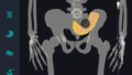
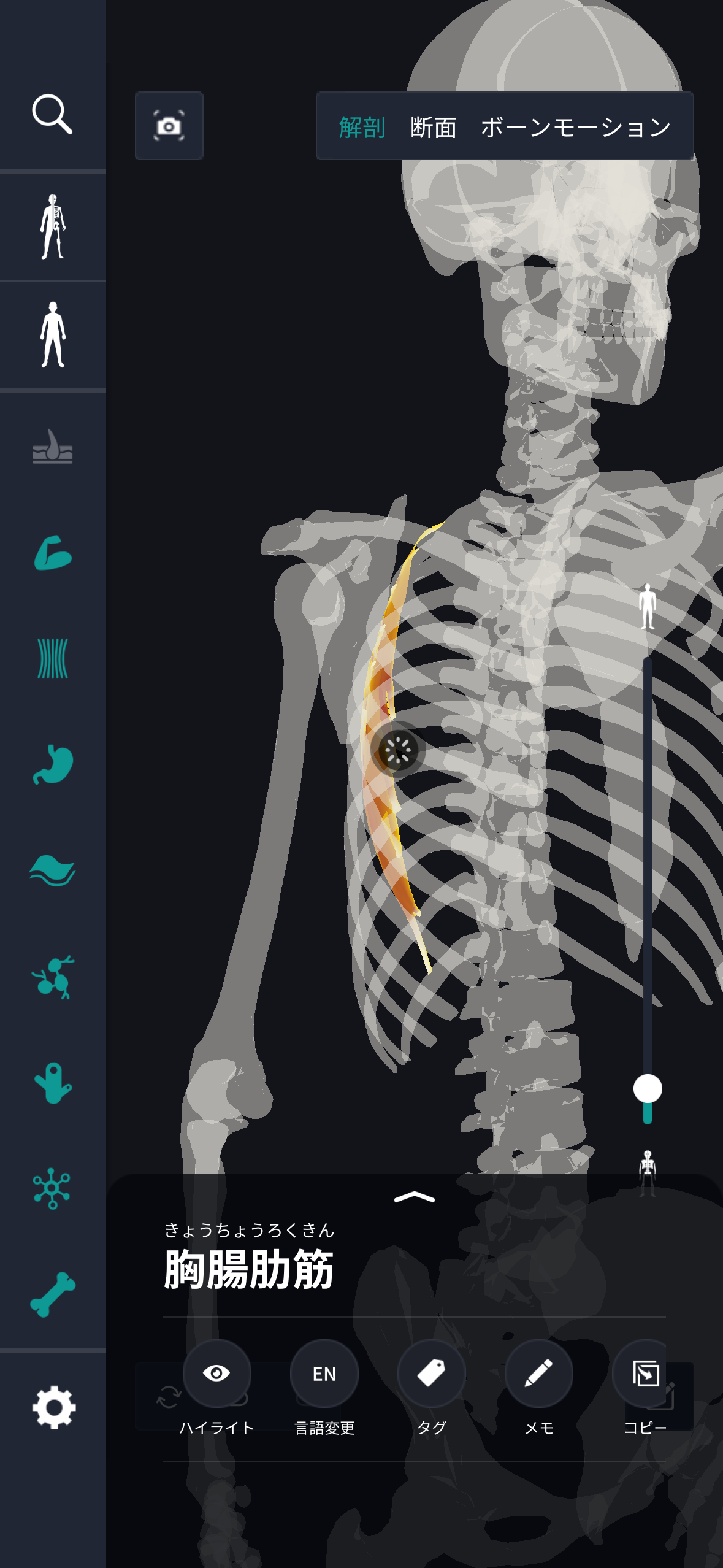
In the anatomy application, you can view a selection of anatomy 3D models. In this model, there are various observation methods such as surfaces, cross-sections, and nervous systems. This time, I'll explain using an anatomy application.
About pleural muscles
Study points
The location and structure of the pleural muscles
The diaphragm is the main muscle that separates the chest from the abdomen. Positionally, it is attached to the lower part of the ribs and covers the entire rib cage like an X shape. Specifically, it starts at the back of the sternum, the lower ribs, and the front of the lumbar spine, and converges to the center of the central tendon to form a dome-shaped shape. This dome-shaped shape is usually curved upward, but the shape changes as you breathe. When inhaling, the diaphragm contracts, the dome-shaped shape becomes flat, and the chest cavity expands, increasing the space to take in air. When exhaling, the muscles relax and return to the dome shape again. Structurally, the entire diaphragm can be divided into three major parts. First, the sternum portion is attached to the lower end of the sternum, and then the rib portion is connected to the inner surface of the lower ribs. Also, the lumbar part extends forward of the spine and supports the connection from the dorsal side. Additionally, the diaphragm has several openings, which are essential when considering important structures. The esophageal hiatus passes through the esophagus and the vagus nerve, and the aorta and thoracic duct pass through the aortic hiatus. Also, the inferior vena cava carries blood from the abdomen to the heart through the venous hiatus vena cava. As described above, the diaphragm is not just a muscle; it is a dynamic wall that closely supports breathing, blood circulation, and digestive movements. It can be seen that its location and structure play a central role in various functions in the human body.
The role and function of the pleural muscles
The diaphragm plays a role as the main respiratory muscle and controls the breathing process by moving the lungs. Specifically, when the diaphragm contracts, its dome-shaped part falls downward, and as the volume of the chest cavity increases, the lungs expand, and outside air is taken in. This action is called “breathing” and is essential in basic breathing mechanisms. When inhaling, the diaphragm flattens and the ribs expand slightly outward, reducing intrathoracic pressure. This negative pressure draws outside air into the lungs and promotes oxygen uptake. Conversely, when the diaphragm relaxes, it returns to a dome shape, and air is pushed out by squeezing the lungs, and “exhalation” is performed. In addition to breathing, the diaphragm also contributes to core stability. In particular, it functions as an inner muscle when lifting heavy objects or maintaining body posture, and holds internal organs in the correct position. In addition, it also plays an important role in movements such as laughing, coughing, and sneezing. This is because the diaphragm controls exhalation and supports intentional breathing movements by assisting vocal cord movement. It also supports the operation of the digestive system by increasing abdominal pressure, and also has the effect of helping with bowel movements. In this way, the diaphragm is not simply a muscle that helps breathing; it is an important entity that flexibly responds to and works with various body functions.
English notation for pleural muscle
The diaphragm is expressed as “diaphragm” in English. This word is derived from the ancient Greek word “delta α (diáphragma)” and means “separation” or “partition.” It can be seen from this name that since ancient Greek times, it was recognized that this muscle plays a role in dividing different parts of the body. “diaphragm” in English generally indicates something that structurally or functionally acts as a partition or boundary, and in addition to diaphragms in the human body, parts used as diaphragms to produce sound, for example, speakers of audio devices are also sometimes called “diaphragms.” Furthermore, this term is also used for devices that adjust the lens aperture of a camera, and it is a term widely used in the fields of technology and acoustics. The diaphragm in our body is an important structure that separates the thoracic cavity from the abdominal cavity, and the versatile usage of this word in English shows its applicability to other fields. On a daily basis, we hear “dialysis” in the context of medicine and physiology, but in order to understand its function as a concept, it is important how this structure forms various partitions and has various roles. Thus, the English notation for diaphragm not only indicates one body part, but also has a wide range of meanings reflecting the scope of use, history, and structural characteristics of the word.
How to study human anatomy
I will explain specific study methods using human anatomy applications.
Check your past learning history and practice repeatedly
Here are the steps to check your anatomy learning history and practice iteratively effectively.
1. Check your learning history in the app
Reviewing your learning history with the application is an important step in effectively advancing anatomy learning. First, launch the app and go to the learning history section from the main menu. Many anatomy apps are designed to show your progress in the form of graphs and lists, so you can visually check which parts you've learned about and how much time you've spent.
By using this data, you can understand which areas you have strengths in and where you need to spend more time and effort. We also recommend using a dedicated tag or notebook function to mark areas you are particularly weak at or where you need to relearn. Regularly checking your learning history and looking back on past learning content will lead to efficient review and deepening understanding.
2.Make a plan for iterative learning
Making an efficient repetitive learning plan based on learning history is extremely effective in promoting knowledge retention. First, identify weak points and areas where you need to relearn. Next, arrange these study items into a weekly or monthly calendar and create a specific study schedule. By proceeding in a planned manner, you can learn each part evenly and avoid packing in a large amount of information at once.
Using a task management app or digital calendar to set study reminders is effective. Also, it's important to have the flexibility to regularly review progress and revise plans as needed. By having goals and proceeding with your studies in a planned manner, you can efficiently acquire anatomical knowledge.
3.Use 3D features to learn visually
By utilizing the 3D function, learning anatomy is easier to understand visually. The 3D model shows the structure of the human body three-dimensionally, and each part can be observed in detail. This makes it possible to intuitively grasp positional relationships between deep muscles and organs that are difficult to capture in a planar view. For example, you can learn even the smallest details by rotating specific muscles and bones and zooming in and out.
Also, there are many apps that have the function of displaying cross-sectional views of each part using a 3D model, which is useful for deepening understanding of internal structures. This diversity of visual information helps with memory retention and improves immediate responsiveness in tests and practice situations. By utilizing the 3D function and learning visually, you can learn anatomy knowledge more deeply and efficiently.
Use the memo function concretely

Test your learning regularly in the form of quizzes
Regularly testing what you've learned in a quiz format is a very effective way to anchor your anatomy knowledge. Quiz-style tests help you objectively grasp your level of understanding and areas you lack while repeating knowledge.
For example, by using a learning app to conduct quizzes every specific period, you can reconfirm what you've learned and strengthen your memory. There are a wide range of quiz formats, such as multiple choice questions, fill-in-the-blank questions, and short answer questions, and each helps understanding from a different angle and develops the ability to utilize various types of knowledge.
Get feedback
If possible, get feedback from other learners and experts. It helps you find your own gaps in understanding and areas for improvement. You can also keep yourself motivated to learn by regularly testing yourself. Feeling a sense of accomplishment and progress increases motivation for continuous learning.
summary
This time, I explained how to study “thoracic bowel muscles” using an application!
Thank you for reading this far.
I would be happy if reading this article helped you learn about anatomy.
Learning is a long, never-ending journey, but I sincerely wish you all the best. Let's continue to study together and work hard for the national exam!
Please look forward to the next blog.