beginning
In this article, I will explain effective study methods, starting with knowledge of specialized parts in human anatomy.
In human anatomy, it is necessary not only to memorize the names of various organs, muscles, and bones, but also to remember where they are located in the body. Therefore, it is necessary to learn as efficiently as possible.
I hope you can read this article and use the app to deepen your understanding even a little bit.
Now, I will explain the details about the “interosseous cuneus ligament” and how to study human anatomy.
teamLab Body Pro Free Download
A 3D anatomy app that shows all the structures of the human body
Download teamLab Body Pro here!

What is the interosseous cuneiform ligament?
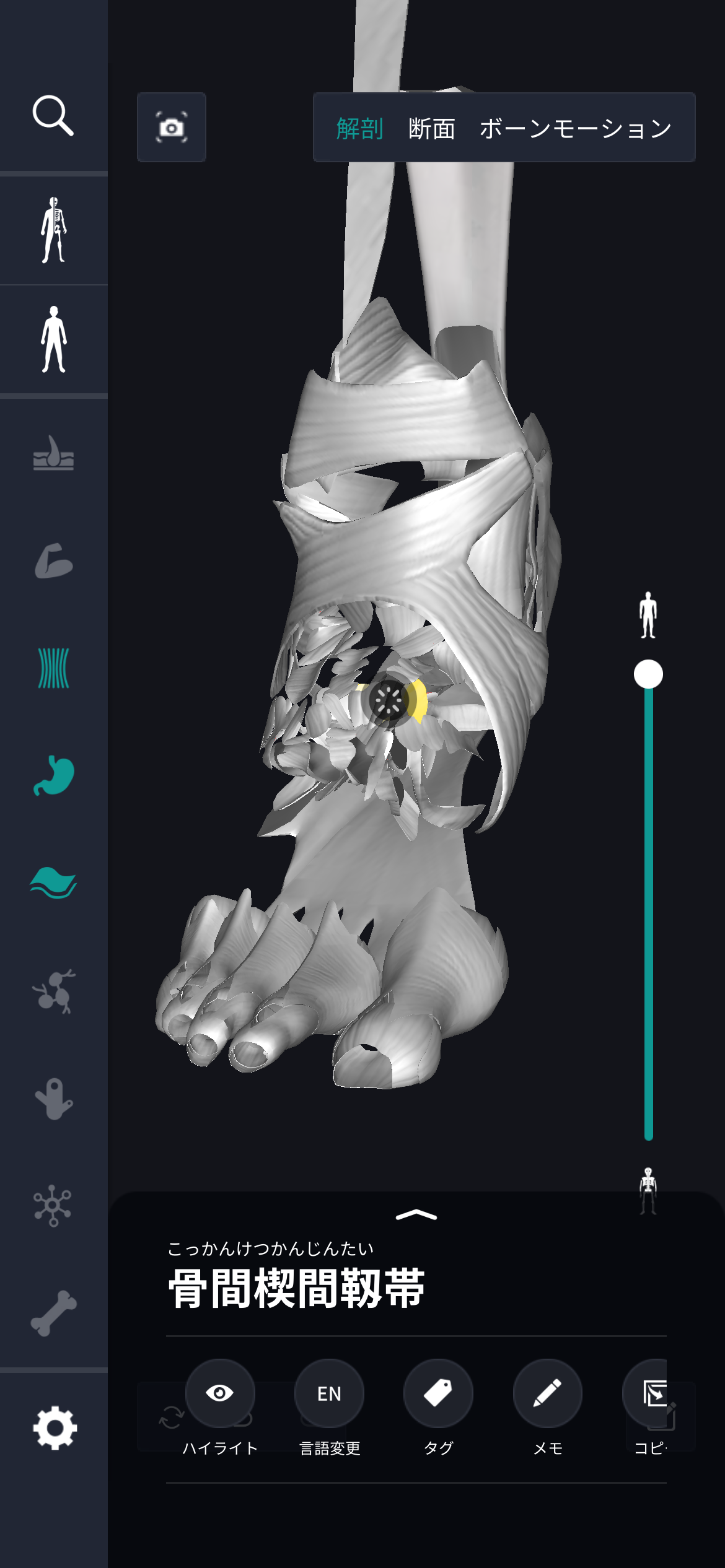
In the anatomy application, you can view a selection of anatomy 3D models. In this model, there are various observation methods such as surfaces, cross-sections, and nervous systems. This time, I'll explain using an anatomy application.
About interosseous cuneiform ligament

The interosseous intercuneal ligament is one of the important ligaments in the foot structure and mainly supports metatarsal stability. Many bones are involved in the foot, and they have a role in connecting the cuneiform bone, which is the core part, and the scaphoid bone. The bone structure of the foot is complex; multiple bones form joints, and ligaments connect between them. The ligaments themselves are organized with tough fibers and play a role in reinforcing joints and limiting excessive movement. It absorbs shock when the foot lands on the ground, and also plays a mechanical role in smoothly advancing walking movements. The normal function of this ligament is very important not only for daily walking activities, but also for movements such as running. Also, the strength and condition of ligaments can be affected by age and physical condition, and exercise and activity may be affected. Therefore, understanding the structure of the foot, including ligaments, is key to maintaining health without putting a strain on the foot. It is recommended to avoid obsessive movements and balance the body through general foot care.
Study points
Location and structure of interosseous cuneiform ligament
By understanding the location and structure of the interosseous cuneus ligament, we can find contributions to foot stability and function. This ligament is located on the inside of the foot between the cuneiform bones and even between the scaphoid bones. The foot skeleton is a complex combination of many small bones, which are tightly connected by ligaments. Ligaments are composed of extremely flexible and tough fibrous tissue and are intended to strengthen and stabilize joints. The flexibility and strength of the interosseous cuneiform ligament efficiently absorbs forces generated during foot movement and provides stability during walking. Ligaments also play a role in supporting normal walking patterns by maintaining the arrangement of foot bones. The firm structure of this ligament forms a support base for the foot to move safely, and functional advantages can also be obtained in everyday movements and sports activities. A basic understanding of the location and structure of ligaments is useful for maintaining foot health. This information can help you be aware of the structure of your foot and be able to take proper care without neglecting it.
The role and function of the interosseous cuneiform ligament
The role and function of the interosseous cuneus ligament is very important in foot movement and stability. This ligament maintains the arch of the foot and performs a mechanical function to properly distribute impact when the foot is stressed. Ligaments are composed of firm fibrous tissue, which binds the bones of the foot to each other and contributes to joint stabilization. In particular, when walking or running, the foot absorbs the force when it lands on the ground and prepares preparations for a smooth transition to the next movement. This ligament between the cuneiform and scaphoid bones provides structural support for the foot and relieves stress generated during repetitive movements. This allows the legs to move efficiently and maintain body balance. Ligament damage or excessive load may interfere with foot movement and affect the movement of the whole body. Therefore, proper care and attention are important to protect ligaments and maintain overall foot health in everyday life. Understanding the role and function of interosseous cuneiform ligaments is essential in considering continued foot health.
English notation for interosseous cuneiform ligament
The interosseous cuneiform ligament is called “interosseous intercuneiform ligament” in English. This name specifically describes the location and role of specific ligaments in the foot. “interosseous” has the meaning of “interosseous” and specifically indicates that it is located between the bones of the foot. “intercuneiform” means “cuneiform,” and “cuneiform” refers to cuneiform bone. Cuneiform bones are composed of multiple small bones located in the metapod of the foot, and this name is used because there are ligaments between them. The word ligament is “ligament,” and it has the meaning of “something that binds”, and its tissue binds bones and plays a role in maintaining joint stability. These English expressions help you understand the structure of the foot and provide basic information to know where ligaments are located and what purpose they have. Based on such explanations, you can be aware of foot movement and function, and use it as knowledge to promote general health. By avoiding specialized content and using this information, you will deepen your understanding of how the foot works.
How to study human anatomy
I will explain specific study methods using human anatomy applications.
Check your past learning history and practice repeatedly
Here are the steps to check your anatomy learning history and practice iteratively effectively.
1. Check your learning history in the app
Reviewing your learning history with the application is an important step in effectively advancing anatomy learning. First, launch the app and go to the learning history section from the main menu. Many anatomy apps are designed to show your progress in the form of graphs and lists, so you can visually check which parts you've learned about and how much time you've spent.
By using this data, you can understand which areas you have strengths in and where you need to spend more time and effort. We also recommend using a dedicated tag or notebook function to mark areas you are particularly weak at or where you need to relearn. Regularly checking your learning history and looking back on past learning content will lead to efficient review and deepening understanding.
2.Make a plan for iterative learning
Making an efficient repetitive learning plan based on learning history is extremely effective in promoting knowledge retention. First, identify weak points and areas where you need to relearn. Next, arrange these study items into a weekly or monthly calendar and create a specific study schedule. By proceeding in a planned manner, you can learn each part evenly and avoid packing in a large amount of information at once.
Using a task management app or digital calendar to set study reminders is effective. Also, it's important to have the flexibility to regularly review progress and revise plans as needed. By having goals and proceeding with your studies in a planned manner, you can efficiently acquire anatomical knowledge.
3.Use 3D features to learn visually
By utilizing the 3D function, learning anatomy is easier to understand visually. The 3D model shows the structure of the human body three-dimensionally, and each part can be observed in detail. This makes it possible to intuitively grasp positional relationships between deep muscles and organs that are difficult to capture in a planar view. For example, you can learn even the smallest details by rotating specific muscles and bones and zooming in and out.
Also, there are many apps that have the function of displaying cross-sectional views of each part using a 3D model, which is useful for deepening understanding of internal structures. This diversity of visual information helps with memory retention and improves immediate responsiveness in tests and practice situations. By utilizing the 3D function and learning visually, you can learn anatomy knowledge more deeply and efficiently.
Use the memo function concretely

Make notes so you don't forget the things and points you've noticed while studying. The memo function can be used for different purposes, such as inputting text, saving images, and writing memos. Tag your notes to make them easier to review later.
Test your learning regularly in the form of quizzes
Regularly testing what you've learned in a quiz format is a very effective way to anchor your anatomy knowledge. Quiz-style tests help you objectively grasp your level of understanding and areas you lack while repeating knowledge.
For example, by using a learning app to conduct quizzes every specific period, you can reconfirm what you've learned and strengthen your memory. There are a wide range of quiz formats, such as multiple choice questions, fill-in-the-blank questions, and short answer questions, and each helps understanding from a different angle and develops the ability to utilize various types of knowledge.
Get feedback
If possible, get feedback from other learners and experts. It helps you find your own gaps in understanding and areas for improvement. You can also keep yourself motivated to learn by regularly testing yourself. Feeling a sense of accomplishment and progress increases motivation for continuous learning.
summary
This time, I explained how to study about the “interosseous cuneiform ligament” using an application!
Thank you for reading this far.
I would be happy if reading this article helped you learn about anatomy.
Learning is a long, never-ending journey, but I sincerely wish you all the best. Let's continue to study together and work hard for the national exam!
Please look forward to the next blog.