Where is the cerebellum? Location, vermis and hemispheres, peduncles and function
Where is the cerebellum?
Is the cerebellum part of the cerebrum? No. Cup the back of your head just above the hairline: the cerebellum lies deep inside, near the floor of the skull. It tucks under the back half of the cerebrum and sits against the back of the brainstem.
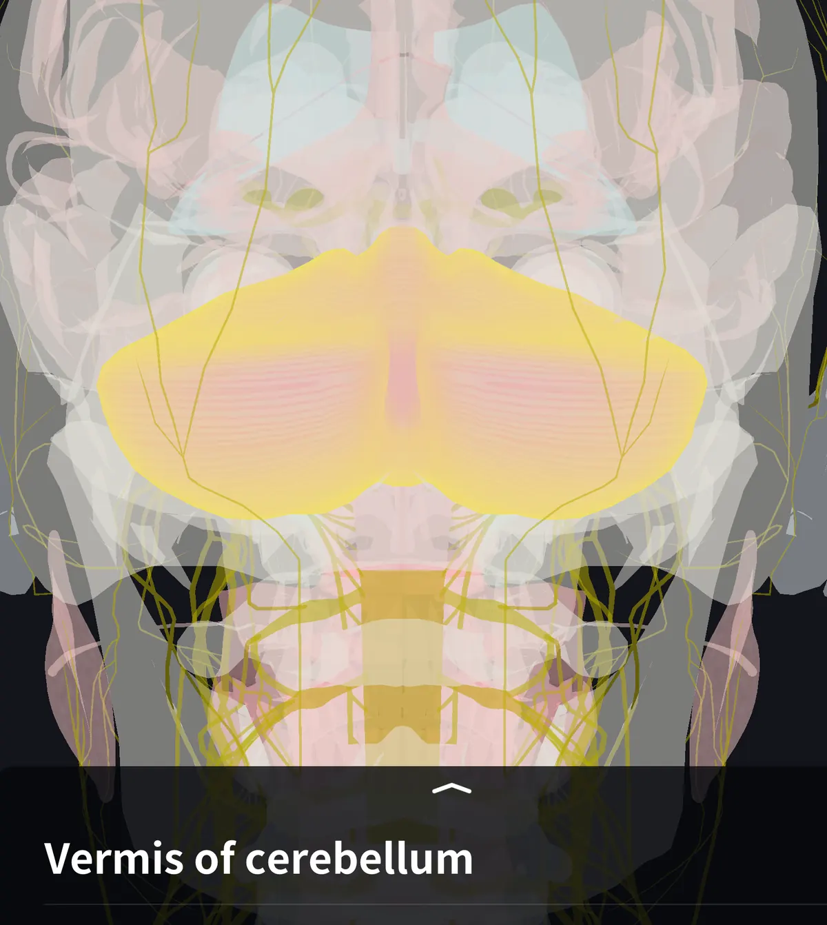
The inside floor of the skull steps down in three levels. The cerebellum fills the lowest and most posterior one, the posterior cranial fossa. Above it stretches the tentorium cerebelli, a sheet of dura mater on which the occipital lobes rest. An adult cerebellum weighs about 130–150 g.
That is only about a tenth of the brain's weight, yet more than half of all the brain's neurons are here, mostly tiny granule cells packed into the deep layer of the cerebellar cortex.
Relation to the cerebrum and brainstem
Picture the brain cut down the midline. From front to back you see the cerebrum, the brainstem (midbrain, pons and medulla) below it, and the cerebellum behind the brainstem. Between the cerebellum and the brainstem lies the fourth ventricle, a space filled with cerebrospinal fluid; the cerebellum forms its roof.
The cerebellum never touches the cerebrum directly: the tentorium always lies between them. Signals from the cerebrum reach it by way of the pons.
Shape and parts
Seen from behind, the cerebellum looks like a butterfly with open wings. The narrow middle strip is the vermis (Latin for "worm", after its ridged, worm-like look) and the large wings on either side are the cerebellar hemispheres.
・Vermis: the narrow midline part joining the two hemispheres
・Cerebellar hemispheres: the large lateral parts that make up most of the cerebellum
・Flocculonodular lobe: a small part on the front of the underside, made of the nodule of the vermis and the paired flocculi
The surface is creased by fine, almost parallel grooves, with narrow ridges between them called folia. Two deep fissures divide it into lobes: the primary fissure separates the anterior and posterior lobes, and the posterolateral fissure separates the posterior lobe from the flocculonodular lobe. Textbooks differ in how finely they name the smaller lobules, so start with vermis, hemispheres and flocculonodular lobe.
Inside the cerebellum and the peduncles
Cut the cerebellum and you see a thin layer of grey matter, the cerebellar cortex, over branching white matter. The branching pattern is called the arbor vitae, "tree of life".
Deep in the white matter are four pairs of deep cerebellar nuclei: from lateral to medial, the dentate, emboliform, globose and fastigial nuclei. Almost everything the cortex computes passes through these nuclei on its way out to the brainstem and thalamus.
The cerebellum is attached to the brainstem by three pairs of cerebellar peduncles. Their names match the level of the brainstem they join.
・Superior cerebellar peduncle: joins the midbrain; carries mainly fibres leaving the cerebellum
・Middle cerebellar peduncle: joins the pons; the thickest, carrying input from the cerebrum relayed in the pons
・Inferior cerebellar peduncle: joins the medulla; carries input from the spinal cord and the vestibular system of the inner ear
What the cerebellum does
The cerebellum does not start movements. It compares the commands sent by the cerebrum with information about how the body is actually moving, from muscles, joints and the inner ear, and corrects the difference. That is why your fingers stop in the right place when you reach for a cup.
By function and connections it is usually divided into three parts, each named after the structure it mainly talks to:
・Vestibulocerebellum (flocculonodular lobe): linked with the vestibular system; balance and eye movements
・Spinocerebellum (vermis and medial hemispheres): receives body-position information from the spinal cord; posture and ongoing limb movements
・Cerebrocerebellum (lateral hemispheres): linked with the cerebrum via the pons; planning of skilled movements such as fine finger work
Learning to ride a bicycle or play a fast passage on an instrument also relies on the cerebellum, which gradually stores how to correct errors. How far it is involved in thinking and emotion is still being studied, and textbooks describe this differently.
Name and Japanese term
Cerebellum is the Latin diminutive of cerebrum, "little brain"; the adjective is cerebellar. In Japanese it is 小脳 (shōnō), literally "small brain", and the vermis is 虫部 (chūbu), "worm part".
See it in 3D
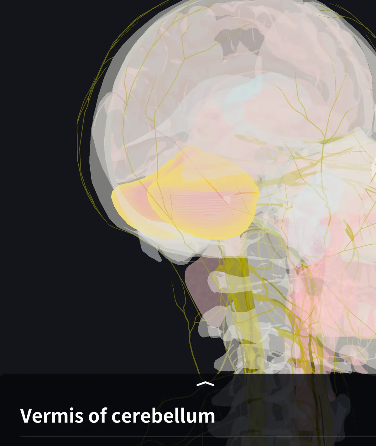
The cerebellum is hidden by the skull and the cerebrum, so flat textbook figures rarely show how it sits behind the brainstem. In the teamLabBody 3D anatomy app you can rotate and zoom the head to view it from behind and below. The vermis and the two hemispheres are separate parts, and the superior, middle and inferior peduncles can each be selected to trace how they meet the midbrain, pons and medulla. With the PREMIUM plan, a midline cross-section shows the cerebellum, brainstem and fourth ventricle together.
FAQ
- Where exactly is the cerebellum?
- At the lower back of the head, deep to the area just above the neck. It lies in the posterior cranial fossa, under the occipital lobes of the cerebrum and behind the brainstem.
- What separates the cerebellum from the cerebrum?
- The tentorium cerebelli, a sheet of dura mater. The occipital lobes rest on top of it and the cerebellum lies beneath it.
- What is the difference between the vermis and the cerebellar hemispheres?
- The vermis is the narrow midline part; the hemispheres are the large parts on either side. The vermis is mainly involved in posture and trunk control, the lateral hemispheres in planning fine limb movements.
Related articles
・Cerebellar hemisphere: the two large lobes in more detail
・Oculomotor nerve: cranial nerve III, leaving the midbrain in front of the cerebellum
・Optic nerve: the visual pathway to the cerebrum