beginning
In this article, I will explain effective study methods, starting with knowledge of specialized parts in human anatomy.
In human anatomy, it is necessary not only to memorize the names of various organs, muscles, and bones, but also to remember where they are located in the body. Therefore, it is necessary to learn as efficiently as possible.
I hope you can read this article and use the app to deepen your understanding even a little bit.
Now, I'll explain the details about the “inferior medial knee vein” and how to study human anatomy.
teamLab Body Pro Free Download
A 3D anatomy app that shows all the structures of the human body
Download teamLab Body Pro here!

What is the inferior medial knee vein?
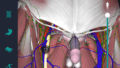
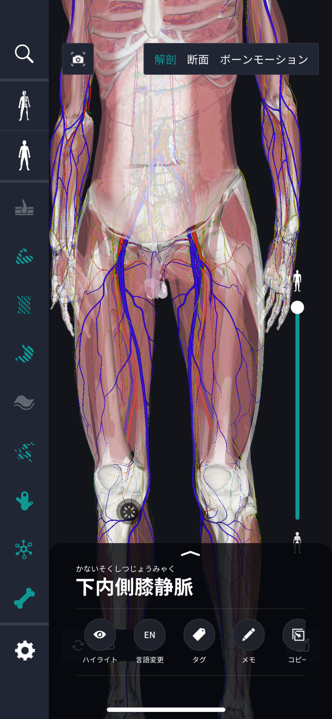
In the anatomy application, you can view a selection of anatomy 3D models. In this model, there are various observation methods such as surfaces, cross-sections, and nervous systems. This time, I'll explain using an anatomy application.
About the inferior medial knee vein

Study points
Location and structure of the inferior medial knee vein
The inferior medial knee vein is a blood vessel that runs inside the knee and is located in the lower part of the popliteal fossa formed by bending the knee joint. This vein flows while collecting blood from the foot before connecting to the femoral vein. As for the structure, it has a flexible tubular shape, and venous valves are arranged. This valve plays a role in preventing blood from flowing back and maintains circulation in one direction. The walls of blood vessels are relatively thin, consist of various internal and external layers, and are designed to maintain stability even when surrounded by muscles. The connection with surrounding tissues, muscles, and connective tissue is very strong, and this adapts to the movement of the knee joint and promotes blood flow. The inferior medial knee vein supports overall circulation by cooperating with other blood vessels and quickly transports blood to the thigh.
The role and function of the inferior medial knee vein
The inferior medial knee vein is part of an important circulatory mechanism responsible for returning blood from the lower extremities to the heart. This vein supports the efficient return of blood to the heart against gravity, especially during exercise or when standing. Since venous valves prevent backflow and ensure unidirectional flow, blood steadily heads to the heart. Also, by being compressed by the contraction of surrounding muscles, it works to push up blood. This process is called muscle pumping and is promoted by walking and exercise. Furthermore, waste products in the blood are eliminated through veins, and normal circulation in the body is maintained, which supports the maintenance of cellular health and function. As an important part of the venous system, the inferior medial knee vein regulates circulation and contributes to maintaining overall body health.
English notation for inferior medial knee vein
The English notation for inferior medial knee vein is “venous medial genicular vein.” This name includes technical terms indicating anatomical locations and characteristics. First, “medial” indicates a “lower” position, and “medial” indicates an “inside” position. “Genicular” is a word meaning “knee,” and “vein” represents “vein.” This name is to express in English the role of a vein that is located inside the knee and collects blood just before it joins the thigh. These notations are widely used in anatomy and medical specialties, and each part accurately describes its function and location. Therefore, although it may seem a bit complicated, it can be said that it is a very useful term for knowing positions and characteristics.
How to study human anatomy
I will explain specific study methods using human anatomy applications.
Check your past learning history and practice repeatedly
Here are the steps to check your anatomy learning history and practice iteratively effectively.
1. Check your learning history in the app
Reviewing your learning history with the application is an important step in effectively advancing anatomy learning. First, launch the app and go to the learning history section from the main menu. Many anatomy apps are designed to show your progress in the form of graphs and lists, so you can visually check which parts you've learned about and how much time you've spent.
By using this data, you can understand which areas you have strengths in and where you need to spend more time and effort. We also recommend using a dedicated tag or notebook function to mark areas you are particularly weak at or where you need to relearn. Regularly checking your learning history and looking back on past learning content will lead to efficient review and deepening understanding.
2.Make a plan for iterative learning
Making an efficient repetitive learning plan based on learning history is extremely effective in promoting knowledge retention. First, identify weak points and areas where you need to relearn. Next, arrange these study items into a weekly or monthly calendar and create a specific study schedule. By proceeding in a planned manner, you can learn each part evenly and avoid packing in a large amount of information at once.
Using a task management app or digital calendar to set study reminders is effective. Also, it's important to have the flexibility to regularly review progress and revise plans as needed. By having goals and proceeding with your studies in a planned manner, you can efficiently acquire anatomical knowledge.
3.Use 3D features to learn visually
By utilizing the 3D function, learning anatomy is easier to understand visually. The 3D model shows the structure of the human body three-dimensionally, and each part can be observed in detail. This makes it possible to intuitively grasp positional relationships between deep muscles and organs that are difficult to capture in a planar view. For example, you can learn even the smallest details by rotating specific muscles and bones and zooming in and out.
Also, there are many apps that have the function of displaying cross-sectional views of each part using a 3D model, which is useful for deepening understanding of internal structures. This diversity of visual information helps with memory retention and improves immediate responsiveness in tests and practice situations. By utilizing the 3D function and learning visually, you can learn anatomy knowledge more deeply and efficiently.
Use the memo function concretely

Test your learning regularly in the form of quizzes
Regularly testing what you've learned in a quiz format is a very effective way to anchor your anatomy knowledge. Quiz-style tests help you objectively grasp your level of understanding and areas you lack while repeating knowledge.
For example, by using a learning app to conduct quizzes every specific period, you can reconfirm what you've learned and strengthen your memory. There are a wide range of quiz formats, such as multiple choice questions, fill-in-the-blank questions, and short answer questions, and each helps understanding from a different angle and develops the ability to utilize various types of knowledge.
Get feedback
If possible, get feedback from other learners and experts. It helps you find your own gaps in understanding and areas for improvement. You can also keep yourself motivated to learn by regularly testing yourself. Feeling a sense of accomplishment and progress increases motivation for continuous learning.
summary
This time, I explained how to study the “inferior medial knee vein” using an application!
Thank you for reading this far.
I would be happy if reading this article helped you learn about anatomy.
Learning is a long, never-ending journey, but I sincerely wish you all the best. Let's continue to study together and work hard for the national exam!
Please look forward to the next blog.