beginning
In this article, I will explain effective study methods, starting with knowledge of specialized parts in human anatomy.
In human anatomy, it is necessary not only to memorize the names of various organs, muscles, and bones, but also to remember where they are located in the body. Therefore, it is necessary to learn as efficiently as possible.
I hope you can read this article and use the app to deepen your understanding even a little bit.
Now, I'll explain the details about the “obturator nerve” and how to study human anatomy.
teamLab Body Pro Free Download
A 3D anatomy app that shows all the structures of the human body
Download teamLab Body Pro here!

What is the obturator nerve?
In the anatomy application, you can view a selection of anatomy 3D models. In this model, there are various observation methods such as surfaces, cross-sections, and nervous systems. This time, I'll explain using an anatomy application.
About the obturator nerve
Study points
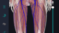
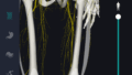
Location and structure of the obturator nerve
The obturator nerve plays an important role due to its location and structure. The obturator nerve starts from the 2nd to 4th nerve roots in the lumbar spine and extends into the pelvis. They exist one by one on the left and right, move from the outside to the inside of the pelvis, and pass through closed holes. This closed pore is a part of the pelvis surrounded by the ischial bone and pubic bone, and the obturator nerve passes through this portal to the lower leg. Nerves move forward in the pelvis and are distributed through the obturator membrane to the adductor muscle group. Structurally, the obturator nerve forms part of the lumbar plexus at the top and branches to the adductor muscle at the bottom, providing motor and sensory control. This nerve is mainly controlled by the spine and is responsible for transmitting sensory and motor information to muscles and skin. Also, since the obturator nerve sends sensory signals to the skin on the inside of the thigh, it can be affected by external forces or changes in position. As a result, when nerve ligation or physical impairment occurs, it may cause paresthesia or muscle weakness on the inside of the thigh.
The role and function of the obturator nerve
The obturator nerve mainly functions as a motor nerve that controls the adductor muscle group of the lower extremities and helps with stability when walking and stopping. This nerve controls the inverted movement of the leg by sending movement commands to muscle groups located on the inside of the thigh in particular. Adduction movements are necessary when pulling the leg inward and are essential for maintaining balance when walking. Also, the obturator nerve acts as a sensory nerve that supplies sensory signals to the skin on the inside of the thigh. This transmits information about touch, pain, and temperature to the brain. The obturator nerve is important for maintaining normal function in sports activities and daily movements, and when impaired, it causes weakening of the adductor muscle group and paresthesia, which affects movement. Therefore, the obturator nerve makes an important contribution to the smooth execution of walking and stability of standing. In particular, when nerve damage or compression occurs, its function declines, leading to loss of balance in gait and paresthesia inside the thigh. These conditions need to be improved through proper diagnosis and treatment.
English notation for obturator nerve
The English notation for obturator nerve is “obturator nerve.” This name comes from the “obturator foramen (obturator foramen)” through which the obturator nerve passes. “obturator” comes from the Latin word “obturare” (close, block) and anatomically indicates the location of nerves in the pelvis. “Nerve” is general English meaning nerve, and it indicates a bundle of fibers that transmit signals as part of the nervous system in the human body. Therefore, the obturator nerve is a standard English notation used to indicate its position and function in anatomy and medicine as an “obturator nerve.” This name is particularly commonly used in academic texts and medical medical certificates. Even in Japan, when the term obturator nerve is expressed in English in medical education or specialized situations, this “obturator nerve” is often used.
How to study human anatomy
I will explain specific study methods using human anatomy applications.
Check your past learning history and practice repeatedly
Here are the steps to check your anatomy learning history and practice iteratively effectively.
1. Check your learning history in the app
Reviewing your learning history with the application is an important step in effectively advancing anatomy learning. First, launch the app and go to the learning history section from the main menu. Many anatomy apps are designed to show your progress in the form of graphs and lists, so you can visually check which parts you've learned about and how much time you've spent.
By using this data, you can understand which areas you have strengths in and where you need to spend more time and effort. We also recommend using a dedicated tag or notebook function to mark areas you are particularly weak at or where you need to relearn. Regularly checking your learning history and looking back on past learning content will lead to efficient review and deepening understanding.
2.Make a plan for iterative learning
Making an efficient repetitive learning plan based on learning history is extremely effective in promoting knowledge retention. First, identify weak points and areas where you need to relearn. Next, arrange these study items into a weekly or monthly calendar and create a specific study schedule. By proceeding in a planned manner, you can learn each part evenly and avoid packing in a large amount of information at once.
Using a task management app or digital calendar to set study reminders is effective. Also, it's important to have the flexibility to regularly review progress and revise plans as needed. By having goals and proceeding with your studies in a planned manner, you can efficiently acquire anatomical knowledge.
3.Use 3D features to learn visually
By utilizing the 3D function, learning anatomy is easier to understand visually. The 3D model shows the structure of the human body three-dimensionally, and each part can be observed in detail. This makes it possible to intuitively grasp positional relationships between deep muscles and organs that are difficult to capture in a planar view. For example, you can learn even the smallest details by rotating specific muscles and bones and zooming in and out.
Also, there are many apps that have the function of displaying cross-sectional views of each part using a 3D model, which is useful for deepening understanding of internal structures. This diversity of visual information helps with memory retention and improves immediate responsiveness in tests and practice situations. By utilizing the 3D function and learning visually, you can learn anatomy knowledge more deeply and efficiently.
Use the memo function concretely

Test your learning regularly in the form of quizzes
Regularly testing what you've learned in a quiz format is a very effective way to anchor your anatomy knowledge. Quiz-style tests help you objectively grasp your level of understanding and areas you lack while repeating knowledge.
For example, by using a learning app to conduct quizzes every specific period, you can reconfirm what you've learned and strengthen your memory. There are a wide range of quiz formats, such as multiple choice questions, fill-in-the-blank questions, and short answer questions, and each helps understanding from a different angle and develops the ability to utilize various types of knowledge.
Get feedback
If possible, get feedback from other learners and experts. It helps you find your own gaps in understanding and areas for improvement. You can also keep yourself motivated to learn by regularly testing yourself. Feeling a sense of accomplishment and progress increases motivation for continuous learning.
summary
This time, I explained how to study “closed nerves” using applications!
Thank you for reading this far.
I would be happy if reading this article helped you learn about anatomy.
Learning is a long, never-ending journey, but I sincerely wish you all the best. Let's continue to study together and work hard for the national exam!
Please look forward to the next blog.