beginning
In this article, I will explain effective study methods, starting with knowledge of specialized parts in human anatomy.
In human anatomy, it is necessary not only to memorize the names of various organs, muscles, and bones, but also to remember where they are located in the body. Therefore, it is necessary to learn as efficiently as possible.
I hope you can read this article and use the app to deepen your understanding even a little bit.
Now, I will explain the details about the “lateral collateral ligament of the knee” and how to study human anatomy.
teamLab Body Pro Free Download
A 3D anatomy app that shows all the structures of the human body
Download teamLab Body Pro here!

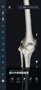
What is the lateral collateral ligament of the knee?
In the anatomy application, you can view a selection of anatomy 3D models. In this model, there are various observation methods such as surfaces, cross-sections, and nervous systems. This time, I'll explain using an anatomy application.
About lateral collateral ligament of knee
Study points
Location and structure of the lateral collateral ligament of the knee
The location and structure of the lateral collateral ligament of the knee forms an important part of the outside of the knee joint. Geographically, it is located on the outside of the knee and is arranged to connect the lateral condyle of the femur to the head of the fibula. This ligament runs throughout the outside of the knee and enhances knee stability in a way that counteracts specific movements and stress. In terms of structure, the lateral collateral ligament of the knee is a thick and relatively short ligament. This ligament consists of fibrous connective tissue and combines elasticity and strength. By running together in a fixed direction, these fibers distribute force from the side against the knee and exhibit the ability to counter it. Also, this ligament cooperates with other muscles, tendons, and ligaments around the knee, and plays a role in maintaining knee stability more comprehensively. Specifically, it supports smooth movement of the knee joint by cooperating with strong muscle groups such as quadriceps femoris. Therefore, a healthy lateral collateral ligament is essential for the overall function and stability of the knee and for comfortable walking in everyday life.
The role and function of the lateral collateral ligament of the knee
The lateral collateral ligament of the knee (lateral collateral ligament) plays a major role in controlling and stabilizing the movement of the knee joint. This ligament is located on the outside of the knee and performs its function, especially by preventing the knee from moving sideways. Specifically, it prevents the knee from moving too much inward or outward, and supports the knee joint to move within the correct range. The lateral collateral ligament has the function of resisting force applied from the side of the knee. Such forces may occur during movements such as walking or running, and the role of ligaments is important. Also, this ligament plays an active role in maintaining knee stability against rapid movements that occur during dynamic activities such as sports. Furthermore, this ligament works in close coordination with other structures in the knee joint. For example, the muscles around the knee support the ligaments and enhance their function. In particular, muscle groups such as the quadriceps femoris and hamstrings cooperate with the lateral collateral ligament in bending and stretching the knee. This cooperative relationship ensures smooth and stable movement throughout the knee. Overall, the lateral collateral ligament of the knee provides the backbone for the proper functioning of the knee in everyday physical activity. As a result, the knee adapts to various movements and can respond efficiently.
English notation for lateral collateral ligament of knee
The English notation for lateral collateral ligament of the knee is “Lateral Collateral Ligament” (LCL). The English name for this ligament succinctly describes its location and function. First, “lateral” means “outside,” and refers to the side away from the center of the body. As this statement indicates, the LCL is located outside the knee joint. Secondly, the word “collateral” means “incidental” or “lateral.” This indicates that LCL is located on the side of the knee joint and plays a role in supporting joint stability. This lateral placement is important to prevent the knee from becoming laterally unstable. Finally, “Ligament” stands for “ligament.” Ligaments are connective tissue that connects bone to bone and provides stability while limiting joint movement. In the case of the lateral collateral ligament of the knee, it plays a role in supporting proper movement of the knee by connecting the femur and fibula. By using this English name, the location and role of the lateral collateral ligament of the knee is widely understood in the international medical field. Even in Japanese, the katakana notation “lateral collateral ligament” is sometimes used, and it is used along with English notation in medical and sports-related literature. This makes it easier for experts to exchange information across national borders, making it possible to gain common understanding.
How to study human anatomy
I will explain specific study methods using human anatomy applications.
Check your past learning history and practice repeatedly
Here are the steps to check your anatomy learning history and practice iteratively effectively.
1. Check your learning history in the app
Reviewing your learning history with the application is an important step in effectively advancing anatomy learning. First, launch the app and go to the learning history section from the main menu. Many anatomy apps are designed to show your progress in the form of graphs and lists, so you can visually check which parts you've learned about and how much time you've spent.
By using this data, you can understand which areas you have strengths in and where you need to spend more time and effort. We also recommend using a dedicated tag or notebook function to mark areas you are particularly weak at or where you need to relearn. Regularly checking your learning history and looking back on past learning content will lead to efficient review and deepening understanding.
2.Make a plan for iterative learning
Making an efficient repetitive learning plan based on learning history is extremely effective in promoting knowledge retention. First, identify weak points and areas where you need to relearn. Next, arrange these study items into a weekly or monthly calendar and create a specific study schedule. By proceeding in a planned manner, you can learn each part evenly and avoid packing in a large amount of information at once.
Using a task management app or digital calendar to set study reminders is effective. Also, it's important to have the flexibility to regularly review progress and revise plans as needed. By having goals and proceeding with your studies in a planned manner, you can efficiently acquire anatomical knowledge.
3.Use 3D features to learn visually
By utilizing the 3D function, learning anatomy is easier to understand visually. The 3D model shows the structure of the human body three-dimensionally, and each part can be observed in detail. This makes it possible to intuitively grasp positional relationships between deep muscles and organs that are difficult to capture in a planar view. For example, you can learn even the smallest details by rotating specific muscles and bones and zooming in and out.
Also, there are many apps that have the function of displaying cross-sectional views of each part using a 3D model, which is useful for deepening understanding of internal structures. This diversity of visual information helps with memory retention and improves immediate responsiveness in tests and practice situations. By utilizing the 3D function and learning visually, you can learn anatomy knowledge more deeply and efficiently.
Use the memo function concretely

Test your learning regularly in the form of quizzes
Regularly testing what you've learned in a quiz format is a very effective way to anchor your anatomy knowledge. Quiz-style tests help you objectively grasp your level of understanding and areas you lack while repeating knowledge.
For example, by using a learning app to conduct quizzes every specific period, you can reconfirm what you've learned and strengthen your memory. There are a wide range of quiz formats, such as multiple choice questions, fill-in-the-blank questions, and short answer questions, and each helps understanding from a different angle and develops the ability to utilize various types of knowledge.
Get feedback
If possible, get feedback from other learners and experts. It helps you find your own gaps in understanding and areas for improvement. You can also keep yourself motivated to learn by regularly testing yourself. Feeling a sense of accomplishment and progress increases motivation for continuous learning.
summary
This time, I explained how to study the “lateral collateral ligament of the knee” using an application!
Thank you for reading this far.
I would be happy if reading this article helped you learn about anatomy.
Learning is a long, never-ending journey, but I sincerely wish you all the best. Let's continue to study together and work hard for the national exam!
Please look forward to the next blog.