beginning
In this article, I will explain effective study methods, starting with knowledge of specialized parts in human anatomy.
In human anatomy, it is necessary not only to memorize the names of various organs, muscles, and bones, but also to remember where they are located in the body. Therefore, it is necessary to learn as efficiently as possible.
I hope you can read this article and use the app to deepen your understanding even a little bit.
Now, I'll explain the details about the “lateral calcaneal ligament” and how to study human anatomy.
teamLab Body Pro Free Download
A 3D anatomy app that shows all the structures of the human body
Download teamLab Body Pro here!

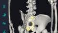
What is lateral calcaneal ligament?
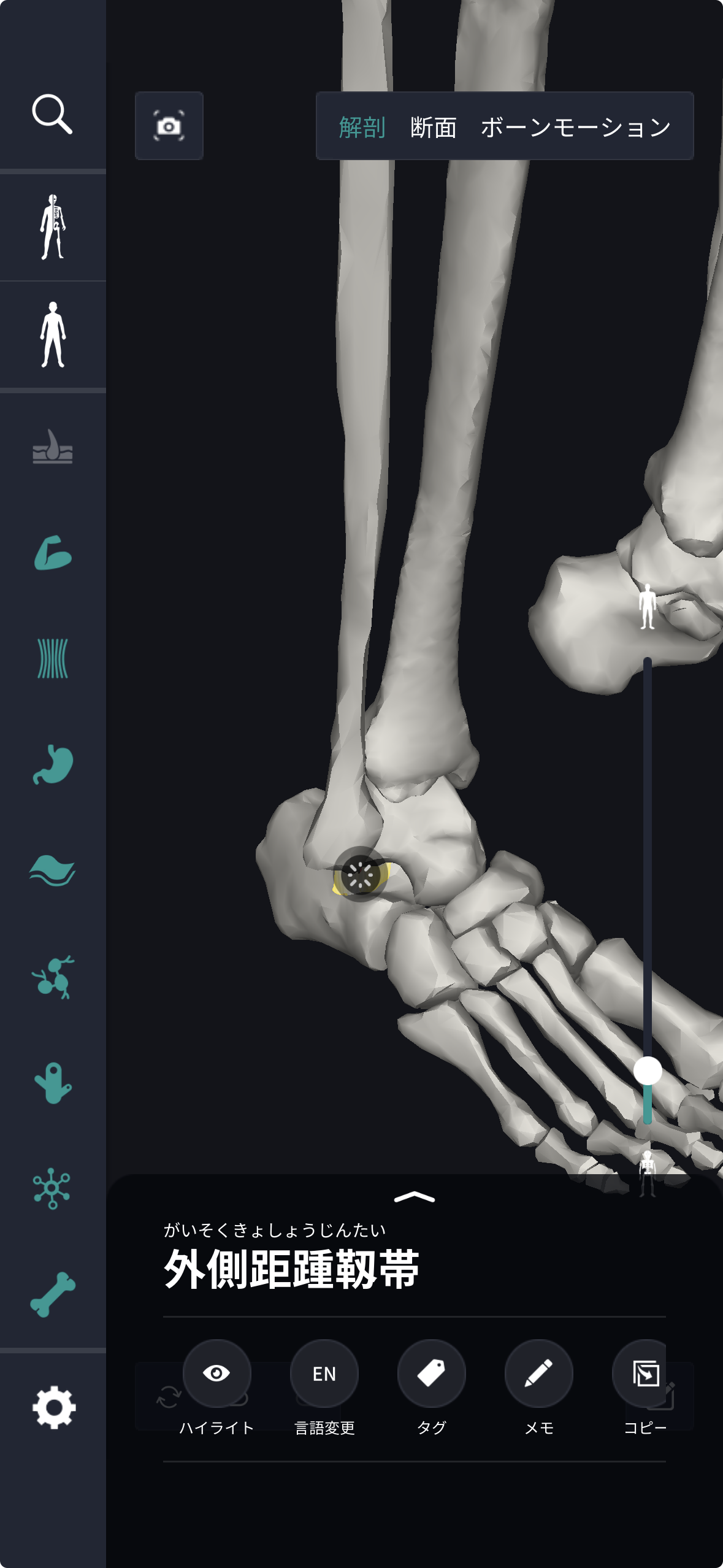
In the anatomy application, you can view a selection of anatomy 3D models. In this model, there are various observation methods such as surfaces, cross-sections, and nervous systems. This time, I'll explain using an anatomy application.
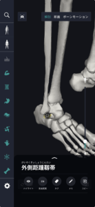
About lateral calcaneal ligament
Study points
Location and structure of lateral calcaneal ligament
The lateral calcaneal ligament is an important ligament located on the outside of the ankle and connects the talus bone to the heel bone. This ligament extends from the back outside of the ankle to the heel, and its arrangement and fiber structure prevent ankle twisting and excessive inward rotation. It looks like a belt and consists of very strong tissue, and can withstand the pressure and stress of everyday life. Structurally, it is formed by bundles of fibers arranged in parallel, thereby providing both high strength and a certain degree of flexibility. This provides a mechanism that stabilizes the ankle while still allowing the delicate movements required for walking and other movements. Furthermore, the lateral calcaneal ligament plays a role in improving overall ankle stability along with other surrounding ligaments and muscles, so it is designed so that this area is not damaged even during sports or strenuous movements. Seen from the outside, these ligaments closely support body movements.
The role and function of the lateral calcaneal ligament
The lateral calcaneal ligament plays an important role in maintaining ankle stability. This ligament functions as a stopper to prevent the ankle from being twisted more than necessary during everyday movements. When we walk, run, or jump, a load is applied to our ankles from various angles, and the lateral calcaneal ligament prevents the ankle from rotating excessively inward during these movements. The elasticity and strength of ligaments live to ensure necessary mobility while absorbing shock applied to the foot. Furthermore, the lateral calcaneal ligament cooperates with other ligaments and muscles in the ankle to promote overall integrated support and movement efficiency. This cooperation ensures ankle movement that can respond flexibly to particularly unstable ground or movements in unexpected directions. Therefore, it can be said that this ligament plays an important role during sports and daily activities. This is because ligaments are structures that distribute the burden on the ankle and allow for a wider range of freedom of movement.
English notation for lateral calcaneal ligament
The name “lateral talocalcaneal ligament,” which is the English notation for lateral calcaneal ligament, is made up of a combination of words that strictly indicate the anatomical position and connection site. First, “lateral” means “outside” far from the center of the body, and the “talocalcaneal” that follows is the “talus (talus),” which is the core of the ankle, and “calcaneus (calcaneus),” which is the heel bone It is an academic compound word connecting the names, and by adding “ligament,” which indicates “ligament,” to it, not only does the spelling itself clearly express that it is literally “a string-like tissue connecting the talus bone to the heel bone on the outside of the ankle,” but it has also been established in English-speaking anatomy as a technical term that symbolizes the stability of the joint composed of these two bones, and beyond a simple symbolic name, where in the body is that tissue exists in words, It is an English notation with a very logical and easy-to-understand etymological structure that contains a specific road map of which bones are connected to which bones.
How to study human anatomy
I will explain specific study methods using human anatomy applications.
Check your past learning history and practice repeatedly
Here are the steps to check your anatomy learning history and practice iteratively effectively.
1. Check your learning history in the app
Reviewing your learning history with the application is an important step in effectively advancing anatomy learning. First, launch the app and go to the learning history section from the main menu. Many anatomy apps are designed to show your progress in the form of graphs and lists, so you can visually check which parts you've learned about and how much time you've spent.
By using this data, you can understand which areas you have strengths in and where you need to spend more time and effort. We also recommend using a dedicated tag or notebook function to mark areas you are particularly weak at or where you need to relearn. Regularly checking your learning history and looking back on past learning content will lead to efficient review and deepening understanding.
2.Make a plan for iterative learning
Making an efficient repetitive learning plan based on learning history is extremely effective in promoting knowledge retention. First, identify weak points and areas where you need to relearn. Next, arrange these study items into a weekly or monthly calendar and create a specific study schedule. By proceeding in a planned manner, you can learn each part evenly and avoid packing in a large amount of information at once.
Using a task management app or digital calendar to set study reminders is effective. Also, it's important to have the flexibility to regularly review progress and revise plans as needed. By having goals and proceeding with your studies in a planned manner, you can efficiently acquire anatomical knowledge.
3.Use 3D features to learn visually
By utilizing the 3D function, learning anatomy is easier to understand visually. The 3D model shows the structure of the human body three-dimensionally, and each part can be observed in detail. This makes it possible to intuitively grasp positional relationships between deep muscles and organs that are difficult to capture in a planar view. For example, you can learn even the smallest details by rotating specific muscles and bones and zooming in and out.
Also, there are many apps that have the function of displaying cross-sectional views of each part using a 3D model, which is useful for deepening understanding of internal structures. This diversity of visual information helps with memory retention and improves immediate responsiveness in tests and practice situations. By utilizing the 3D function and learning visually, you can learn anatomy knowledge more deeply and efficiently.
Use the memo function concretely

Test your learning regularly in the form of quizzes
Regularly testing what you've learned in a quiz format is a very effective way to anchor your anatomy knowledge. Quiz-style tests help you objectively grasp your level of understanding and areas you lack while repeating knowledge.
For example, by using a learning app to conduct quizzes every specific period, you can reconfirm what you've learned and strengthen your memory. There are a wide range of quiz formats, such as multiple choice questions, fill-in-the-blank questions, and short answer questions, and each helps understanding from a different angle and develops the ability to utilize various types of knowledge.
Get feedback
If possible, get feedback from other learners and experts. It helps you find your own gaps in understanding and areas for improvement. You can also keep yourself motivated to learn by regularly testing yourself. Feeling a sense of accomplishment and progress increases motivation for continuous learning.
summary
This time, I explained how to study the “lateral calcaneal ligament” using an application!
Thank you for reading this far.
I would be happy if reading this article helped you learn about anatomy.
Learning is a long, never-ending journey, but I sincerely wish you all the best. Let's continue to study together and work hard for the national exam!
Please look forward to the next blog.