beginning
In this article, I will explain effective study methods, starting with knowledge of specialized parts in human anatomy. In human anatomy, it is necessary not only to memorize the names of various organs, muscles, and bones, but also to remember where they are located in the body. Therefore, it is necessary to learn as efficiently as possible. I hope you can read this article and use the app to deepen your understanding even a little bit. Now, I'll explain the details about the “posterior sacroiliac ligament” and how to study human anatomy.teamLab Body Pro Free Download
A 3D anatomy app that shows all the structures of the human body
Download teamLab Body Pro here!

What is posterior sacroiliac ligament?
In the anatomy application, you can view a selection of anatomy 3D models. In this model, there are various observation methods such as surfaces, cross-sections, and nervous systems. This time, I'll explain using an anatomy application.About posterior sacroiliac ligament
Study points
Location and structure of posterior sacroiliac ligament
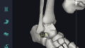
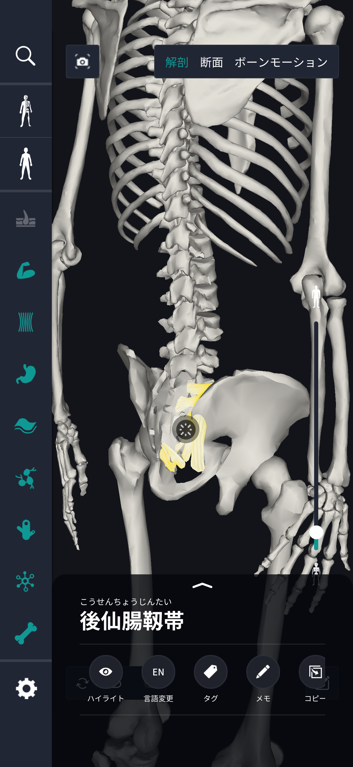
The location and structure of the posterior sacroiliac ligament will be explained. This ligament is located in the pelvic part of the human body and has the role of connecting the sacrum to the iliac bone, that is, the triangular bone (sacrum) below the lumbar spine and the iliac bones on both sides. The posterior sacroiliac ligament is fixed to the back of the pelvis, has a wide shape, and extends to cover the posterior surface of the sacroiliac joint. This ligament is located between the sacrum and iliac bone to maintain pelvic stability. Structurally, the posterior sacroiliac ligament consists of very strong fibrous connective tissue. These fibers are tightly intertwined to maintain their strength. Furthermore, the ligament tissue is also flexible, which not only supports the movement of the human body but also enables moderate movement. In this way, the structure of the pelvis and the function of the posterior sacroiliac ligament are harmonized, helping the stability and movement of the body. Specific clinical details are avoided here, but this is an overview of the basic position and structure of the posterior sacroiliac ligament.The role and function of the posterior sacroiliac ligament
I will explain the role and function of the posterior sacroiliac ligament. This ligament is extremely important for ensuring stability in the human body. Since the posterior sacroiliac ligament supports the pelvis, the center of gravity of the human body is maintained properly, and the weight of the body can be distributed well. As a result, it helps the body stabilize while standing, walking, and other activities. Specifically, by firmly connecting the sacrum and iliac bone, the position of these bones is fixed, and resistance to body movement is increased. This function also has the effect of protecting pelvic structures from excessive movement and impact. Furthermore, the posterior sacroiliac ligament also plays a role in supporting body movements. The flexibility of the ligament tissue supports lower back movement and range of motion when the body bends or twists. Thus, the balance between strength and flexibility of ligaments enables various body movements and provides structural support for the human body. Due to these effects, the posterior sacroiliac ligament functions as an essential structure for the human body, but clinical details are not discussed here.English notation for posterior sacroiliac ligament
The posterior sacroiliac ligament is expressed as “posterior sacroiliac ligament” in English. The name of this ligament indicates the “sacrum” (sacrum) and “iliac bone” (iliac bone) connected at both ends. “Posterior” means “posterior,” indicating that this ligament is located on the posterior surface of the sacroiliac joint. This notation makes it easier to understand the specific location of ligaments and structures related to them. In the medical field, English notation is widely used around the world, facilitates communication between experts, and is useful for international clinical research and education. Also, English names representing each structure of the body are essential for deepening specialized understanding of human anatomy. By using English notation together in a form that complements understanding of Japanese notation, expansion of medical knowledge and deepening of understanding are promoted. This is the basic idea behind the English notation for posterior sacroiliac ligament. Clinical details are omitted here, but the significance of this notation is also important in the broader medical community.How to study human anatomy
I will explain specific study methods using human anatomy applications.Check your past learning history and practice repeatedly
Here are the steps to check your anatomy learning history and practice iteratively effectively. 1. Check your learning history in the appReviewing your learning history with the application is an important step in effectively advancing anatomy learning. First, launch the app and go to the learning history section from the main menu. Many anatomy apps are designed to show your progress in the form of graphs and lists, so you can visually check which parts you've learned about and how much time you've spent.
By using this data, you can understand which areas you have strengths in and where you need to spend more time and effort. We also recommend using a dedicated tag or notebook function to mark areas you are particularly weak at or where you need to relearn. Regularly checking your learning history and looking back on past learning content will lead to efficient review and deepening understanding.
Use the memo function concretely