beginning
In this article, I will explain effective study methods, starting with knowledge of specialized parts in human anatomy.
In human anatomy, it is necessary not only to memorize the names of various organs, muscles, and bones, but also to remember where they are located in the body. Therefore, it is necessary to learn as efficiently as possible.
I hope you can read this article and use the app to deepen your understanding even a little bit.
Now, I will explain the contents of the “oculomotor nerve” and how to study human anatomy.
teamLab Body Pro Free Download
A 3D anatomy app that shows all the structures of the human body
Download teamLab Body Pro here!

What is the oculomotor nerve?
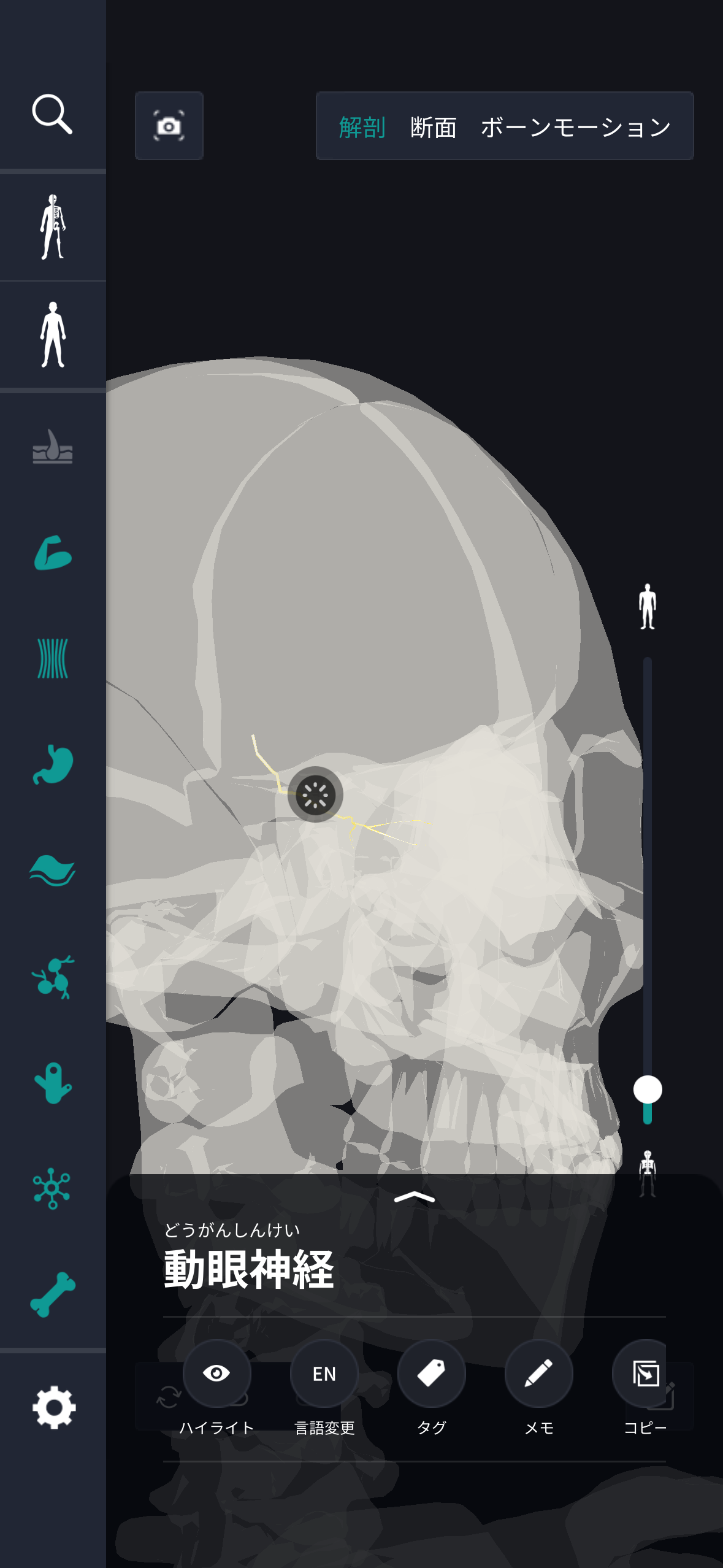
In the anatomy application, you can view a selection of anatomy 3D models. In this model, there are various observation methods such as surfaces, cross-sections, and nervous systems. This time, I'll explain using an anatomy application.
About oculomotor nerve

The oculomotor nerve is one of the cranial nerves and plays an important role in facial muscles and eye movements. This nerve, which originates from the center of the brain, controls eye movement and is responsible for functions such as adjusting gaze, constricting pupils, and opening the eyelids. The oculomotor nerve is particularly important in the parasympathic nervous system and helps process information related to vision. This nerve is divided into 3 main branches, and each transmits information to different eye muscles. This allows the eye to move in all directions. Furthermore, the oculomotor nerve adjusts the amount of light through pupil reflection and also has the function of optimizing the visual experience. The activity of the oculomotor nerve is often not consciously noticed in everyday life, but it exists as an essential element for adjusting vision and eye movements, and when it functions properly, visual information can be collected and recognized smoothly. Thus, the oculomotor nerve is recognized as an important component in the smooth operation of the visual system.
Study points
The location and structure of the oculomotor nerve
The oculomotor nerve is one of the cranial nerves and is sometimes called the III cranial nerve. This nerve starts near the front of the midbrain, passes through the cavernous sinus, and travels within the skull. The oculomotor nerve is located in the dural space at the base of the skull and passes close to the crosstalk and pituitary gland. Structurally, it mainly consists of a mixture of motor nerve fibers and parasympathetic nerve fibers, and each plays a different role. Motor fibers control several muscles involved in eye movement, specifically the medial rectus muscle, superior rectus muscle, inferior rectus muscle, and inferior oblique muscle. This makes it possible for the eyeballs to move in various directions. Parasympathetic fibers have the function of constricting the pupil and help adjust the amount of light and adjust the focal length. Furthermore, the oculomotor nerve also innervates the levator eyelid muscle, and by lifting the eyelids, it contributes to the movement of opening the eyes. The structure and location of this nerve plays an important role in eye movement and function as part of the visual system. It enables eye movement and is the foundation that supports accurate acquisition and processing of visual information.
The role and function of the oculomotor nerve
The role and function of the oculomotor nerve is mainly related to eye movement control and pupil adjustment. This nerve transmits motor commands from the brain and plays an important function for smoothly advancing visual experiences. The motor component of the oculomotor nerve maintains an appropriate direction of sight by sending signals to the muscles that move the eyeball, making it possible to collect complex visual information. This makes it possible to accurately capture the position and movement of objects, and provides essential functions for daily life and environmental recognition. Furthermore, this nerve contains parasympathetic fibers, which contribute to the pupil's function of regulating the amount of light. By supporting the adjustment effect of constricting pupils in bright places and dilating in dark places, the amount of light is managed appropriately and the quality of vision is maintained. Furthermore, the oculomotor nerve is involved in the movement of opening the eyelid via the levator eyelid muscle, and helps visual input to be performed smoothly without being blocked. This series of functions plays a central role in ensuring visual clarity and achieving coordinated eye movement. In this way, the oculomotor nerve has become an important nerve function that supports the integration and efficient processing of visual information.
English notation for oculomotor nerve
The oculomotor nerve in English is expressed as “Oculomotor Nerve.” “Oculo” comes from the Latin word for eye, and “motor” is a word meaning movement. Thus, as can be seen from the English notation, it has been shown that the oculomotor nerve is a nerve involved in eye movement. Since it is the third cranial nerve, it is also called the “Third Cranial Nerve.” This nerve, like other cranial nerves, uses words reflecting its main function and role according to its name. Language, even in medical jargon, functions as a means of communication and helps share scientific understanding between different cultures. The English notation for oculomotor nerve helps share and unify knowledge in the international field of medicine, and is an important element for health care professionals from various countries to cooperate to develop research and treatment methods. Language is more than just a communication tool; it also plays a role as a means of sharing knowledge. In the fields of medicine and science, it is important that experts from different countries and regions have a common understanding through effective communication and cooperate to continue making progress. Since many biological concepts, not limited to oculomotor nerves, are expressed and understood in such a way, extensive cooperation across countries is possible.
How to study human anatomy
I will explain specific study methods using human anatomy applications.
Check your past learning history and practice repeatedly
Here are the steps to check your anatomy learning history and practice iteratively effectively.
1. Check your learning history in the app
Reviewing your learning history with the application is an important step in effectively advancing anatomy learning. First, launch the app and go to the learning history section from the main menu. Many anatomy apps are designed to show your progress in the form of graphs and lists, so you can visually check which parts you've learned about and how much time you've spent.
By using this data, you can understand which areas you have strengths in and where you need to spend more time and effort. We also recommend using a dedicated tag or notebook function to mark areas you are particularly weak at or where you need to relearn. Regularly checking your learning history and looking back on past learning content will lead to efficient review and deepening understanding.
2.Make a plan for iterative learning
Making an efficient repetitive learning plan based on learning history is extremely effective in promoting knowledge retention. First, identify weak points and areas where you need to relearn. Next, arrange these study items into a weekly or monthly calendar and create a specific study schedule. By proceeding in a planned manner, you can learn each part evenly and avoid packing in a large amount of information at once.
Using a task management app or digital calendar to set study reminders is effective. Also, it's important to have the flexibility to regularly review progress and revise plans as needed. By having goals and proceeding with your studies in a planned manner, you can efficiently acquire anatomical knowledge.
3.Use 3D features to learn visually
By utilizing the 3D function, learning anatomy is easier to understand visually. The 3D model shows the structure of the human body three-dimensionally, and each part can be observed in detail. This makes it possible to intuitively grasp positional relationships between deep muscles and organs that are difficult to capture in a planar view. For example, you can learn even the smallest details by rotating specific muscles and bones and zooming in and out.
Also, there are many apps that have the function of displaying cross-sectional views of each part using a 3D model, which is useful for deepening understanding of internal structures. This diversity of visual information helps with memory retention and improves immediate responsiveness in tests and practice situations. By utilizing the 3D function and learning visually, you can learn anatomy knowledge more deeply and efficiently.
Use the memo function concretely

Make notes so you don't forget the things and points you've noticed while studying. The memo function can be used for different purposes, such as inputting text, saving images, and writing memos. Tag your notes to make them easier to review later.
Test your learning regularly in the form of quizzes
Regularly testing what you've learned in a quiz format is a very effective way to anchor your anatomy knowledge. Quiz-style tests help you objectively grasp your level of understanding and areas you lack while repeating knowledge.
For example, by using a learning app to conduct quizzes every specific period, you can reconfirm what you've learned and strengthen your memory. There are a wide range of quiz formats, such as multiple choice questions, fill-in-the-blank questions, and short answer questions, and each helps understanding from a different angle and develops the ability to utilize various types of knowledge.
Get feedback
If possible, get feedback from other learners and experts. It helps you find your own gaps in understanding and areas for improvement. You can also keep yourself motivated to learn by regularly testing yourself. Feeling a sense of accomplishment and progress increases motivation for continuous learning.
summary
This time, I explained how to study “oculomotor nerves” using an application!
Thank you for reading this far.
I would be happy if reading this article helped you learn about anatomy.
Learning is a long, never-ending journey, but I sincerely wish you all the best. Let's continue to study together and work hard for the national exam!
Please look forward to the next blog.