beginning
In this article, I will explain effective study methods, starting with knowledge of specialized parts in human anatomy.
In human anatomy, it is necessary not only to memorize the names of various organs, muscles, and bones, but also to remember where they are located in the body. Therefore, it is necessary to learn as efficiently as possible.
I hope you can read this article and use the app to deepen your understanding even a little bit.
Now, I will explain the details about the “medial collateral ligament of the knee” and how to study human anatomy.
teamLab Body Pro Free Download
A 3D anatomy app that shows all the structures of the human body
Download teamLab Body Pro here!

What is the medial collateral ligament of the knee?
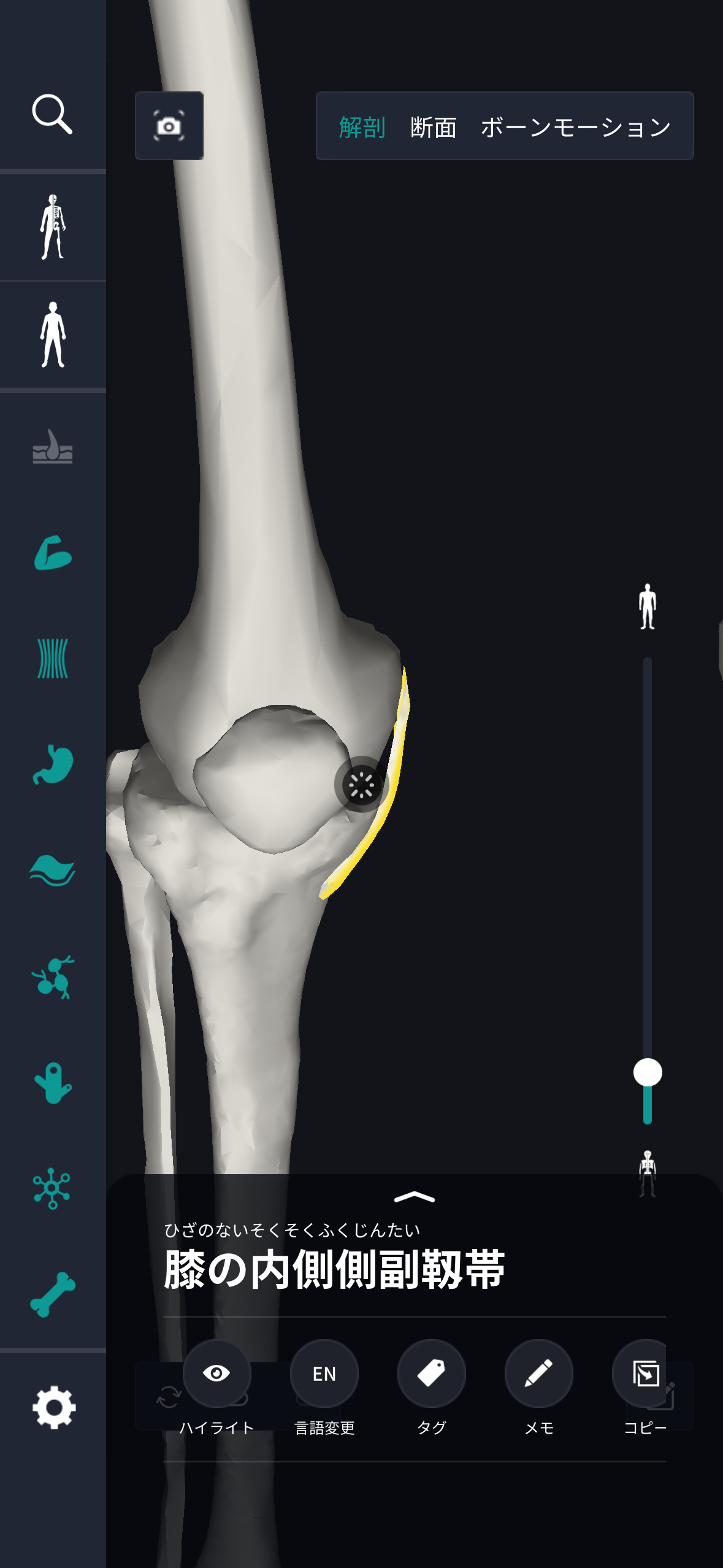
In the anatomy application, you can view a selection of anatomy 3D models. In this model, there are various observation methods such as surfaces, cross-sections, and nervous systems. This time, I'll explain using an anatomy application.
About medial collateral ligament of knee

Study points
Location and structure of the medial collateral ligament of the knee
The medial collateral ligament of the knee is an important component of the knee joint located on the middle side of the body. It is a ligament about 10 cm long that extends inward from the femur to the tibia, and is located along the inside of the knee. This ligament plays a role in preventing the knee from sliding too far sideways or breaking inward. In terms of structure, the medial collateral ligament of the knee is made of strong fibrous connective tissue and has a dense and elastic structure. This provides firm support for knee movements and allows them to withstand movement and pressure in various directions. Also, the flexibility of this ligament enables supple movement when bending and stretching the knee, and plays a role in making the entire body move smoothly. The medial collateral ligament of the knee works in cooperation with other ligaments and muscles to achieve a sense of unity throughout the knee joint. Above all, it supports not only static stability (stability when the knee is not moving), but also dynamic stability (stability while the knee is moving), so it is very important for everyday movement and exercise. For this reason, it is essential to correctly recognize the location and role of the medial collateral ligament when understanding the structure of the knee.
The role and function of the medial collateral ligament of the knee
The medial collateral ligament of the knee supports normal knee function and plays an important role in ensuring movement and stability. The main function of this ligament is to provide stability to the inside of the knee. In particular, it supports the knee so that it does not bend too much inward, and ensures proper movement. This limits movement of the knee in the lateral direction and allows proper movement of the knee to be maintained. Furthermore, the medial collateral ligament protects the knee during various activities. For example, it works to relieve stress on the knee and prevent damage during sports or daily activities. Knee stability is important when the entire body moves, and it provides support to maintain knee balance, especially during sudden changes of direction, jumping, or running. This ligament also works with other structures. When bending and stretching the knee, it cooperates with muscles and other ligaments to enable complex movement of the knee joint. It also fixes the joints when the knee is in an upright position and plays a role in ensuring posture stability. These functions improve physical comfort and functionality, and are the foundation for supporting daily activities.
English notation for medial collateral ligament of knee
The English notation for medial collateral ligament of the knee is “medial collateral ligament.” This term refers to one of the ligaments that make up the knee joint. “Medium” has the meaning of “inside” and indicates the side close to the center line of the human body. “Collateral” is scattered on multiple sides yet has a meaning related to each other, and here it indicates the arrangement of ligaments. Also, “Ligament” means “ligament,” and indicates fibrous tissue that connects bone to bone. Through this name, it becomes clear what role the medial collateral ligament of the knee plays. Since it is located on the inside of the knee, it is suggested that support is provided from within. This allows this ligament to provide restriction and stability to the knee's medial movement. The medical term “medial collateral ligament” facilitates communication between experts and enables accurate information sharing. This notation is frequently used in academic literature and medical documents, and is useful as a means of organizing information related to the knee joint. Also, this name may be useful for the general public to understand medical information.
How to study human anatomy
I will explain specific study methods using human anatomy applications.
Check your past learning history and practice repeatedly
Here are the steps to check your anatomy learning history and practice iteratively effectively.
1. Check your learning history in the app
Reviewing your learning history with the application is an important step in effectively advancing anatomy learning. First, launch the app and go to the learning history section from the main menu. Many anatomy apps are designed to show your progress in the form of graphs and lists, so you can visually check which parts you've learned about and how much time you've spent.
By using this data, you can understand which areas you have strengths in and where you need to spend more time and effort. We also recommend using a dedicated tag or notebook function to mark areas you are particularly weak at or where you need to relearn. Regularly checking your learning history and looking back on past learning content will lead to efficient review and deepening understanding.
2.Make a plan for iterative learning
Making an efficient repetitive learning plan based on learning history is extremely effective in promoting knowledge retention. First, identify weak points and areas where you need to relearn. Next, arrange these study items into a weekly or monthly calendar and create a specific study schedule. By proceeding in a planned manner, you can learn each part evenly and avoid packing in a large amount of information at once.
Using a task management app or digital calendar to set study reminders is effective. Also, it's important to have the flexibility to regularly review progress and revise plans as needed. By having goals and proceeding with your studies in a planned manner, you can efficiently acquire anatomical knowledge.
3.Use 3D features to learn visually
By utilizing the 3D function, learning anatomy is easier to understand visually. The 3D model shows the structure of the human body three-dimensionally, and each part can be observed in detail. This makes it possible to intuitively grasp positional relationships between deep muscles and organs that are difficult to capture in a planar view. For example, you can learn even the smallest details by rotating specific muscles and bones and zooming in and out.
Also, there are many apps that have the function of displaying cross-sectional views of each part using a 3D model, which is useful for deepening understanding of internal structures. This diversity of visual information helps with memory retention and improves immediate responsiveness in tests and practice situations. By utilizing the 3D function and learning visually, you can learn anatomy knowledge more deeply and efficiently.
Use the memo function concretely

Test your learning regularly in the form of quizzes
Regularly testing what you've learned in a quiz format is a very effective way to anchor your anatomy knowledge. Quiz-style tests help you objectively grasp your level of understanding and areas you lack while repeating knowledge.
For example, by using a learning app to conduct quizzes every specific period, you can reconfirm what you've learned and strengthen your memory. There are a wide range of quiz formats, such as multiple choice questions, fill-in-the-blank questions, and short answer questions, and each helps understanding from a different angle and develops the ability to utilize various types of knowledge.
Get feedback
If possible, get feedback from other learners and experts. It helps you find your own gaps in understanding and areas for improvement. You can also keep yourself motivated to learn by regularly testing yourself. Feeling a sense of accomplishment and progress increases motivation for continuous learning.
summary
This time, I explained how to study the “medial collateral ligament of the knee” using an application!
Thank you for reading this far.
I would be happy if reading this article helped you learn about anatomy.
Learning is a long, never-ending journey, but I sincerely wish you all the best. Let's continue to study together and work hard for the national exam!
Please look forward to the next blog.