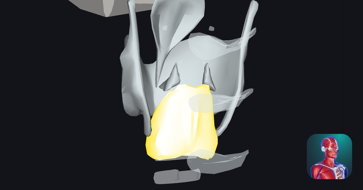
Arytenoid cartilages: shape, processes and how they open and close the glottis
Where are the arytenoid cartilages?
When you speak, your vocal folds come together; when you breathe in, they move apart. What moves them? Two small cartilages holding the back ends of the folds: the arytenoid cartilages.
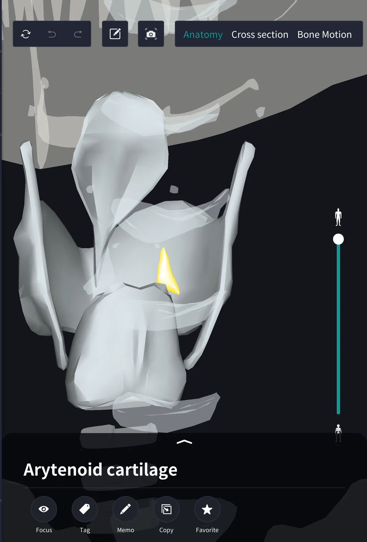
They lie at the back of the larynx, one on each side of the upper border of the tall back plate of the cricoid cartilage. Each is only about the size of a fingertip and cannot be felt from the front of the neck.
A pyramid with two processes
Each arytenoid is a pyramid with a broad base and a pointed apex, on which a tiny corniculate cartilage sits. Two processes project from the base:
・Vocal process: points forward; the back end of the vocal ligament attaches here
・Muscular process: points laterally; the posterior and lateral cricoarytenoid muscles attach here
The vocal folds run from the back of the thyroid cartilage to the vocal processes. The front ends do not move, so the arytenoids do the work of changing the folds.
Rotating and gliding: opening and closing the glottis
At the cricoarytenoid joint each arytenoid rotates about a vertical axis and glides along the upper border of the cricoid lamina. Rotation swings the vocal process in or out, closing or opening the glottis; gliding the two arytenoids together closes its back part.
・Posterior cricoarytenoid: pulls the muscular process back and opens the glottis, the only muscle that does
・Lateral cricoarytenoid: pulls the muscular process forward and closes the glottis
・Transverse and oblique arytenoids: draw the two arytenoids together
Textbooks differ in whether they emphasise rotation or gliding; in practice the two are combined.
Name and terms
Arytenoid comes from Greek words for a ladle or pitcher and "shaped like", said to describe the spout-like shape of the arytenoids and corniculate cartilages together. Related terms: vocal process, muscular process, cricoarytenoid joint. The Latin is cartilago arytenoidea.
See it in the app
Small and hidden behind the thyroid cartilage from the front, the arytenoids are hard to picture. In the 3D anatomy app teamLabBody you can keep only the laryngeal cartilages and rotate to the back to see where they sit on the cricoid and which way the vocal processes point. Cross sections are available with the PREMIUM plan.
FAQ
- Where are the arytenoid cartilages?
- At the back of the larynx, one on each side of the upper border of the cricoid lamina. They cannot be felt from the front of the neck.
- What do the arytenoid cartilages do?
- They hold the back ends of the vocal folds and, by rotating and gliding at the cricoarytenoid joints, open and close the glottis.
- What is the difference between the vocal and muscular processes?
- The vocal process points forward and holds the vocal ligament; the muscular process points laterally and receives the posterior and lateral cricoarytenoid muscles.
Related articles
・Cricoid cartilage: the ring the arytenoids sit on
・Cricoarytenoid ligament: joins the cricoid and arytenoid
・Lateral and posterior cricoarytenoid: close and open the glottis
・Vocal ligament: runs from the arytenoid to the thyroid cartilage
・Larynx: the whole picture