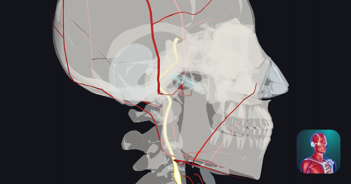
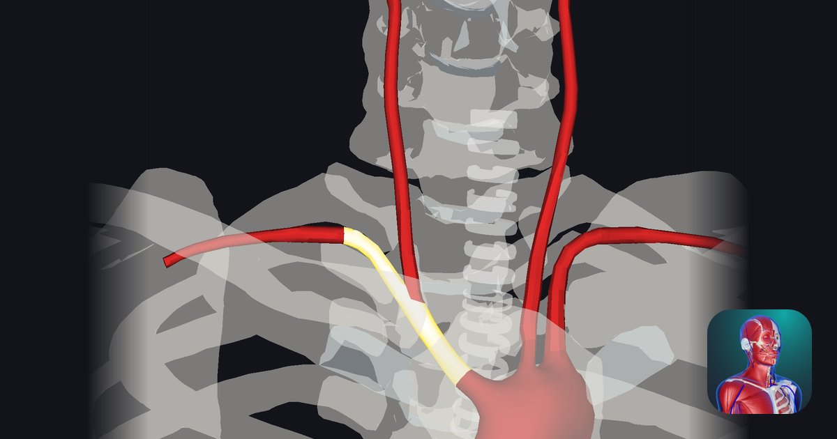
Aorta: course from the heart to the abdomen, segments and arch branches
Where does the aorta run?
Where does blood go first when the heart pumps it out to the body? Into one large artery: the aorta.
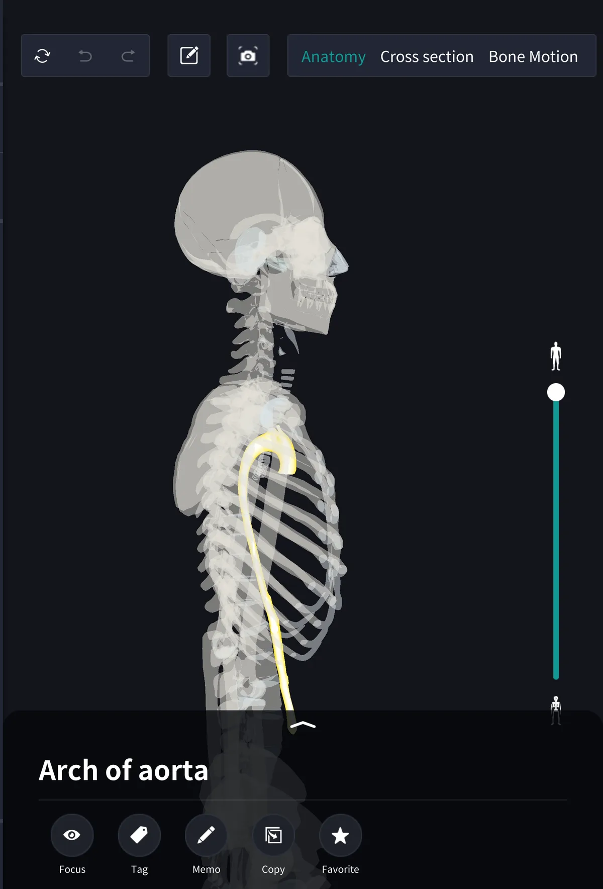
It leaves the left ventricle through the aortic valve, rises, arches backward high in the chest, and then descends in front of the spine to the abdomen, like a walking stick with a curved handle. It is about 2.5–3 cm across where it leaves the heart and narrows further down; the figure varies with the site of measurement, age and individual.
Four segments
・Ascending aorta: the first part, rising from the left ventricle; the coronary arteries arise here
・Aortic arch: curves back and to the left behind the upper sternum
・Thoracic aorta: descends to the left front of the spine, giving off intercostal arteries
・Abdominal aorta: below the diaphragm, along the back wall of the abdomen
The thoracic and abdominal parts together are the descending aorta. teamLabBody names each segment separately; the arch is selected in the view above.
Three branches of the arch
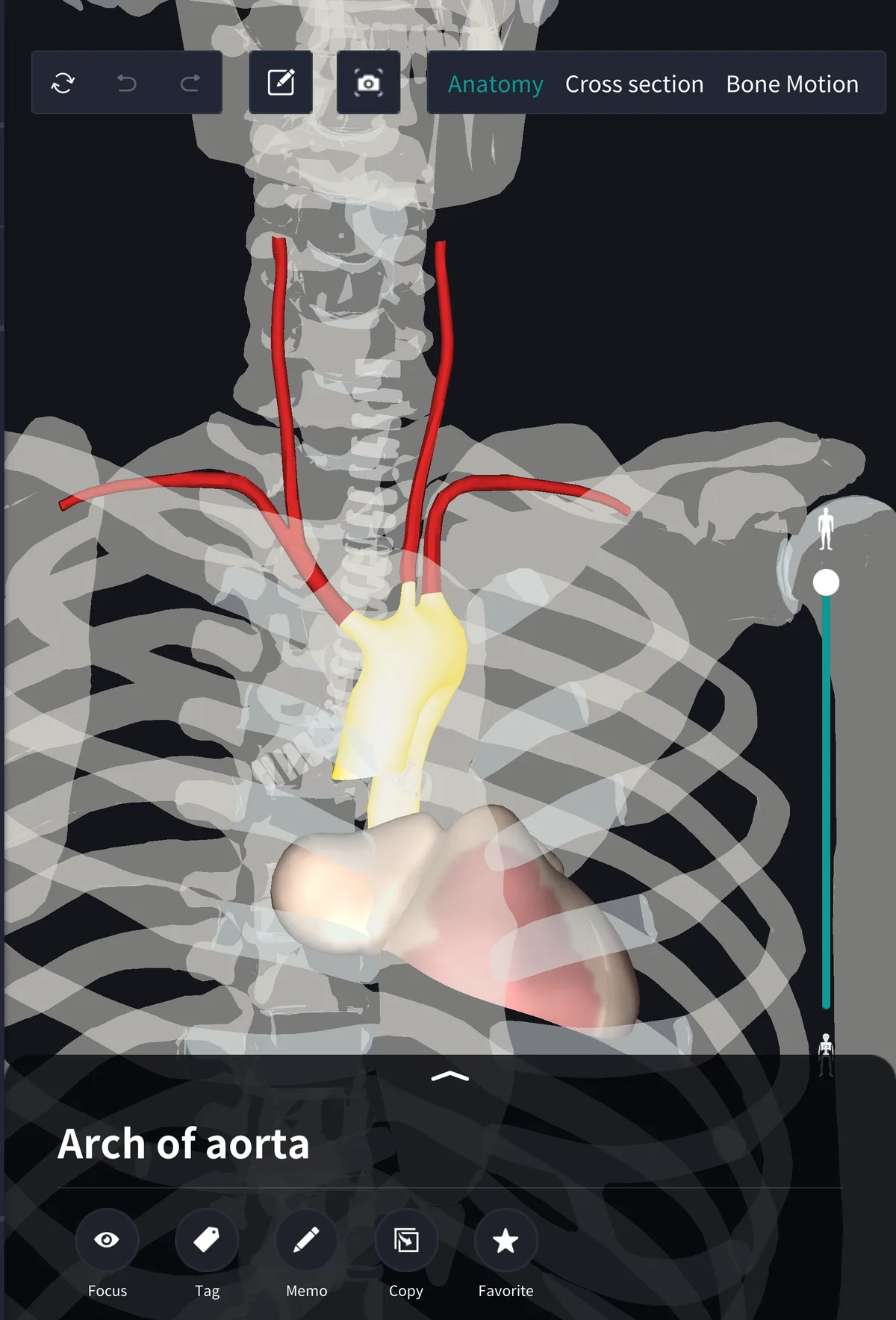
・Brachiocephalic trunk: to the right, soon dividing into the right subclavian and right common carotid arteries
・Left common carotid artery: up the left side of the neck to the head
・Left subclavian artery: to the left arm
On the right the carotid and subclavian share a trunk; on the left they arise from the arch separately, an asymmetry that is easy to mix up. The common carotid in turn divides into the internal and external carotid arteries. Branching patterns vary, and textbooks report different frequencies for variants such as a shared origin of the brachiocephalic and left common carotid arteries.
Through the diaphragm into the abdomen
The thoracic aorta passes through the aortic hiatus at the back of the diaphragm at about T12 and becomes the abdominal aorta. This gives off the coeliac trunk, superior and inferior mesenteric arteries to the gut and the renal arteries, and ends at about L4, a little below the navel, by dividing into the right and left common iliac arteries.
Name and terms
Aorta comes from the Greek aortē, said to derive from a verb meaning to lift. Related terms: ascending aorta, aortic arch, thoracic aorta, abdominal aorta. The Latin is also aorta.
See it in the app
Hidden behind the heart, lungs and gut, the aorta is hard to see whole. In the 3D anatomy app teamLabBody you can make the bones translucent, keep only the aorta, and follow it from the heart to the pelvic brim, then add the heart and arch branches to see where the three branches arise. Cross sections are available with the PREMIUM plan.
FAQ
- Where does the aorta start and end?
- It starts at the left ventricle and runs as ascending aorta, arch, thoracic and abdominal aorta, ending at about L4 by dividing into the common iliac arteries.
- What are the three branches of the aortic arch?
- From right to left: the brachiocephalic trunk, left common carotid artery and left subclavian artery.
- Where does the aorta pass through the diaphragm?
- Through the aortic hiatus at the back of the diaphragm, at about T12.
Related articles
・Left ventricle: pumps blood into the aorta
・Common carotid artery: rises in the neck from the arch branches
・Internal carotid artery: to the brain
・Diaphragm: contains the aortic hiatus
・Vertebral column: the aorta descends in front of it