Internal jugular vein: course from the jugular foramen to the venous angle
Where does the internal jugular vein run?
The internal carotid artery carries blood to the brain. How does that blood get back to the heart? Mainly through the internal jugular vein, deep in the neck.
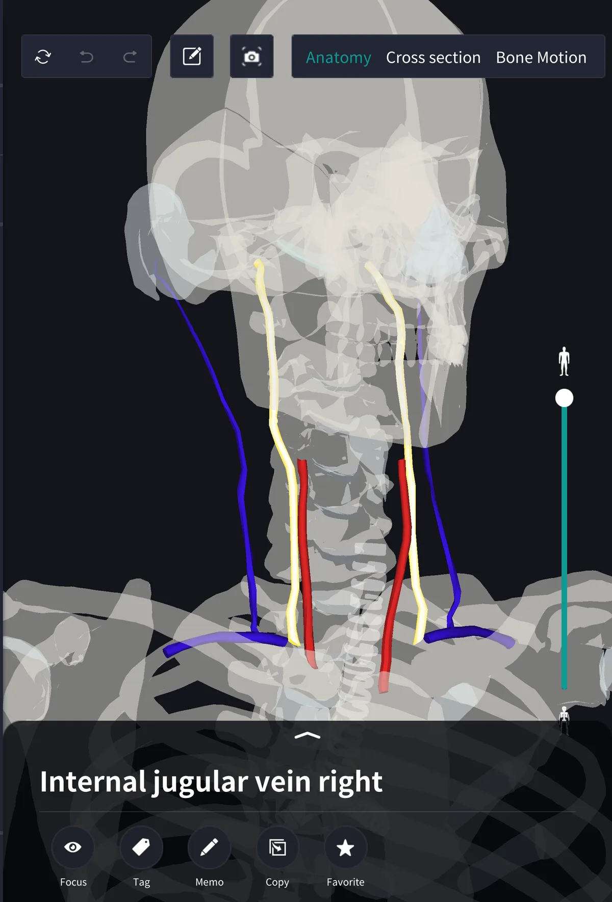
In the view above the internal jugular veins are yellow, the common carotid arteries just medial to them are red, and the blue lines are the superficial external jugular veins. The internal jugular is the largest vein in the neck, usually larger on the right, and runs almost straight from the base of the skull to behind the clavicle.
Beginning: the jugular foramen
It begins at the jugular foramen. Blood from the brain collects in the dural venous sinuses, passes through the S-shaped sigmoid sinus, and leaves the skull here as the internal jugular vein. Its start is slightly dilated (the superior bulb), and there is a similar inferior bulb near its end.
In the neck: the carotid sheath
It runs lateral to the internal carotid artery above and the common carotid artery below, with the vagus nerve between and behind them; all three are enclosed in the carotid sheath, covered by the sternocleidomastoid. On the way down it receives the facial, lingual and thyroid veins, collecting blood from the face and neck as well as the brain.
End: the venous angle and brachiocephalic vein
Behind the medial end of the clavicle it joins the subclavian vein to form the brachiocephalic vein; the two brachiocephalic veins form the superior vena cava, which enters the right atrium. The angle of union is the venous angle, where the thoracic duct (left) and right lymphatic duct (right) return lymph to the blood. The pattern of union varies between people.
Name and terms
Jugular comes from the Latin jugulum, throat or collarbone region. Related terms: external jugular vein, jugular foramen, brachiocephalic vein, venous angle. The Latin is vena jugularis interna.
See it in the app
Overlapped by the sternocleidomastoid and the arteries, the internal jugular vein is hard to place in drawings. In the 3D anatomy app teamLabBody you can show only the vessels, select the vein and rotate to see its relation to the common carotid and its depth compared with the external jugular. Left and right can be selected separately. Cross sections are available with the PREMIUM plan.
FAQ
- Where does the internal jugular vein run?
- From the jugular foramen down the neck lateral to the common carotid artery, to join the subclavian vein behind the medial end of the clavicle.
- How does it differ from the external jugular vein?
- The internal jugular is a large deep vein beside the arteries that also drains the brain; the external jugular is a smaller superficial vein draining mainly the scalp and outer face.
- What is the venous angle?
- The angle where the internal jugular and subclavian veins join; the thoracic duct enters on the left and the right lymphatic duct on the right.
Related articles
・Internal carotid artery: carries blood to the brain
・Common carotid artery: runs medial to the vein
・Vagus nerve: in the carotid sheath with it
・Sternocleidomastoid: covers the carotid sheath