Where is the zygomatic bone? Cheekbone location, zygomatic arch, joints and attached muscles
Where is the zygomatic bone?
When people talk about high cheekbones, this is the bone they mean. Put a finger just below the outer corner of your eye and you will feel a firm ridge. Slide back toward the ear and a thin horizontal bridge of bone runs almost to the ear opening: that is the zygomatic arch.
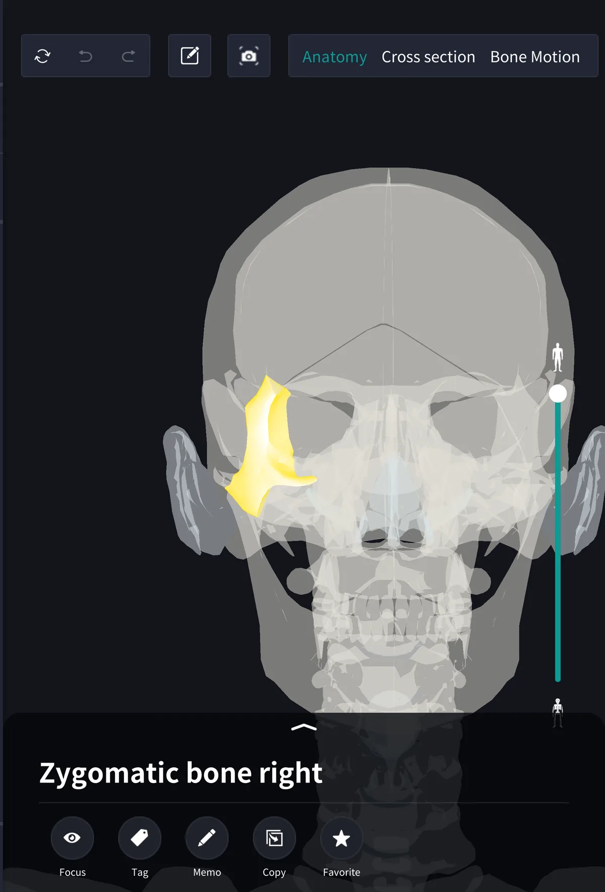
The zygomatic bone is one of the facial bones, one on each side. From the front it sits in the lower outer corner of the orbit; the lateral rim of the orbit and the outer half of its lower rim are made of zygomatic bone.
Four neighbouring bones
・Frontal bone: the frontal process reaches up to meet it below the outer end of the eyebrow
・Temporal bone: the temporal process runs back to join the zygomatic process of the temporal bone, forming the zygomatic arch
・Maxilla: joined in front and medially; the maxilla contains the maxillary sinus
・Sphenoid bone: deep in the orbit it meets the greater wing of the sphenoid to form the lateral orbital wall
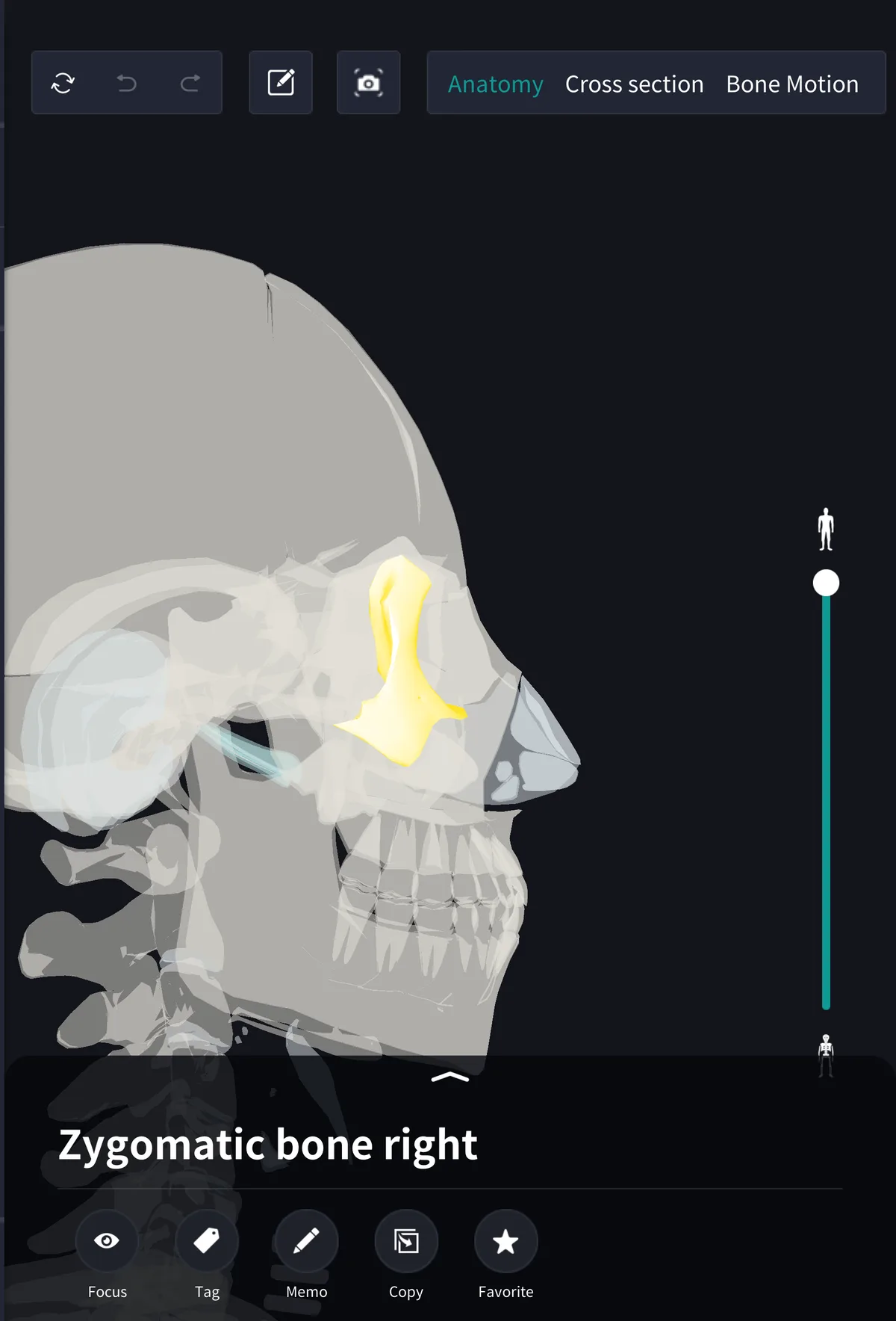
From the side the arch crosses horizontally below the temple. The tendon of the temporalis passes down through the space inside the arch to reach the coronoid process of the mandible, so the arch works as a bony bridge with a tunnel for the muscle underneath.
Surfaces and small foramina
The bone has three surfaces: the lateral (facial) surface, the temporal surface facing the temple, and the orbital surface inside the orbit. The zygomatic nerve, a branch of the maxillary nerve, enters the bone from the orbit and leaves through the zygomaticofacial and zygomaticotemporal foramina to supply the skin of the cheek and temple. The number and size of these foramina vary between individuals.
Muscles attached to the zygomatic bone
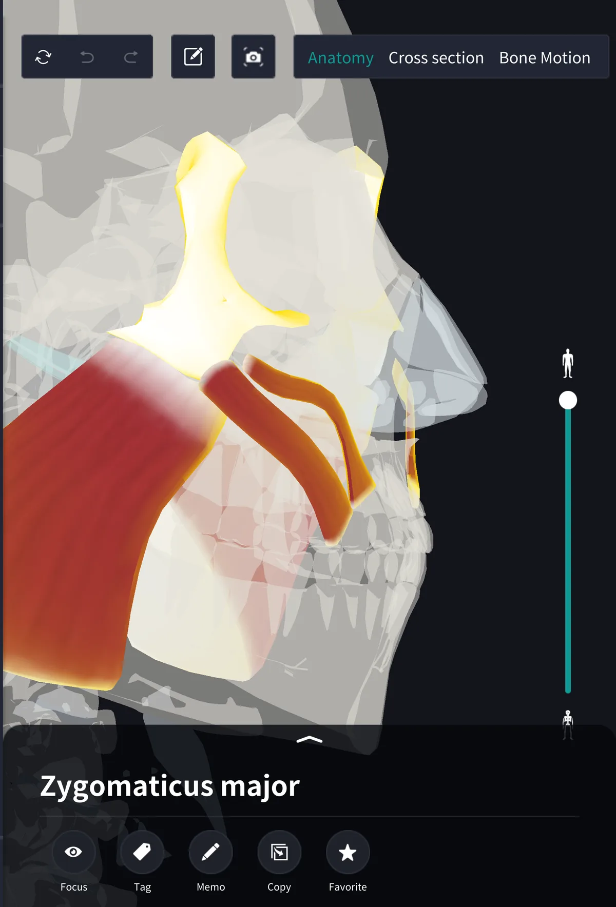
Chewing muscles and smiling muscles both start here. The masseter arises from the lower border and inner surface of the zygomatic arch and runs down to the angle of the mandible; clench your back teeth and you can feel it bulge just below the arch. Its superficial and deep parts arise from the front and back of the arch.
The zygomaticus major and minor arise from the front of the bone. The major runs to the corner of the mouth and the minor to the upper lip, pulling them up and out in a smile. On the temple side, the temporal fascia covering the temporalis attaches to the upper border of the arch.
Name and terms
Zygomatic comes from the Greek word for a yoke, the crossbar joining two oxen, after the way the arch links the temporal bone to the face. Related terms: zygomatic arch, zygomaticofacial foramen, zygomaticus major. The Latin is os zygomaticum.
See it in the app
The zygomatic bone looks completely different from the front and from the side, so a single drawing rarely makes its shape clear. In the 3D anatomy app teamLabBody you can rotate and zoom the head to follow the bone and arch from front, side and oblique views, and turn the muscles back on to see where the masseter and zygomaticus major arise. With the PREMIUM plan, a horizontal cross section shows the gap inside the arch where the temporalis passes.
FAQ
- Where can I feel the zygomatic bone?
- At the high point of the cheek just below the outer corner of the eye. From there you can trace the thin zygomatic arch back to just in front of the ear.
- What is the difference between the zygomatic bone and the zygomatic arch?
- The zygomatic bone is a single bone. The zygomatic arch is a bridge formed by two bones: the temporal process of the zygomatic bone and the zygomatic process of the temporal bone.
- Which muscles attach to the zygomatic bone?
- The masseter arises from the zygomatic arch, and the zygomaticus major and minor from the front of the bone. The temporal fascia attaches to the upper border of the arch.
Related articles
・Masseter: the chewing muscle from the zygomatic arch
・Zygomaticus major and minor: the smiling muscles
・Temporalis: the chewing muscle passing inside the arch
・Maxillary sinus: the air space inside the maxilla