Muscles of mastication: masseter, temporalis and pterygoids, actions and nerve
What are the muscles of mastication?
Clench your back teeth. Two places harden: behind the cheek and at the temple. These are the masseter and temporalis, two of the muscles of mastication.
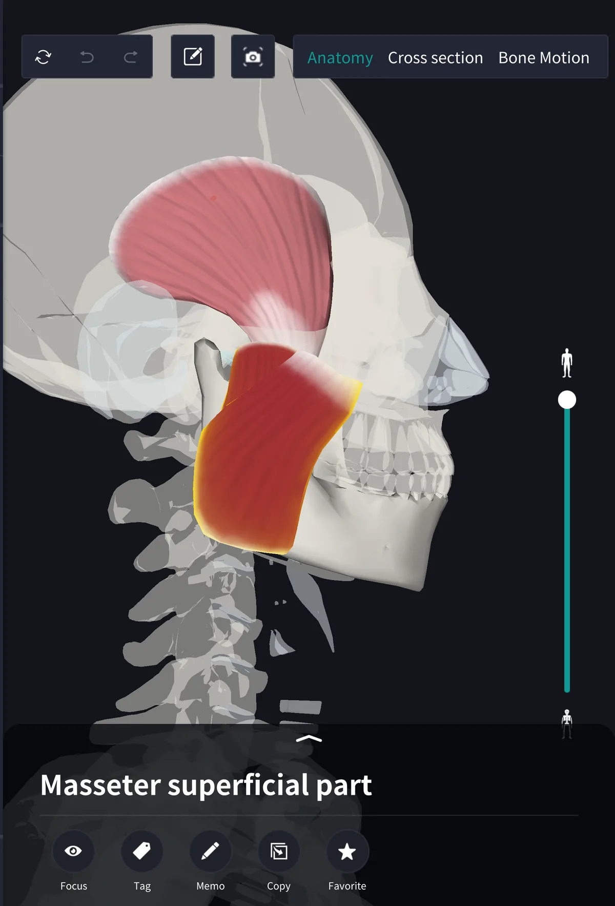
The muscles of mastication move the lower jaw (mandible) when you chew: the masseter, temporalis, medial pterygoid and lateral pterygoid. They are a separate group from the muscles of facial expression, with different attachments and a different nerve. Some textbooks add the suprahyoid muscles such as the digastric as accessory muscles, since they help open the mouth.
Origins and insertions
・Masseter: from the zygomatic arch to the outer surface of the angle of the mandible; superficial and deep parts
・Temporalis: fan-shaped from the temporal fossa, passing inside the zygomatic arch to the coronoid process
・Medial pterygoid: from the medial surface of the lateral pterygoid plate of the sphenoid to the inner surface of the angle
・Lateral pterygoid: from the greater wing of the sphenoid and the lateral surface of the lateral pterygoid plate to the neck of the condylar process and the articular disc of the jaw joint
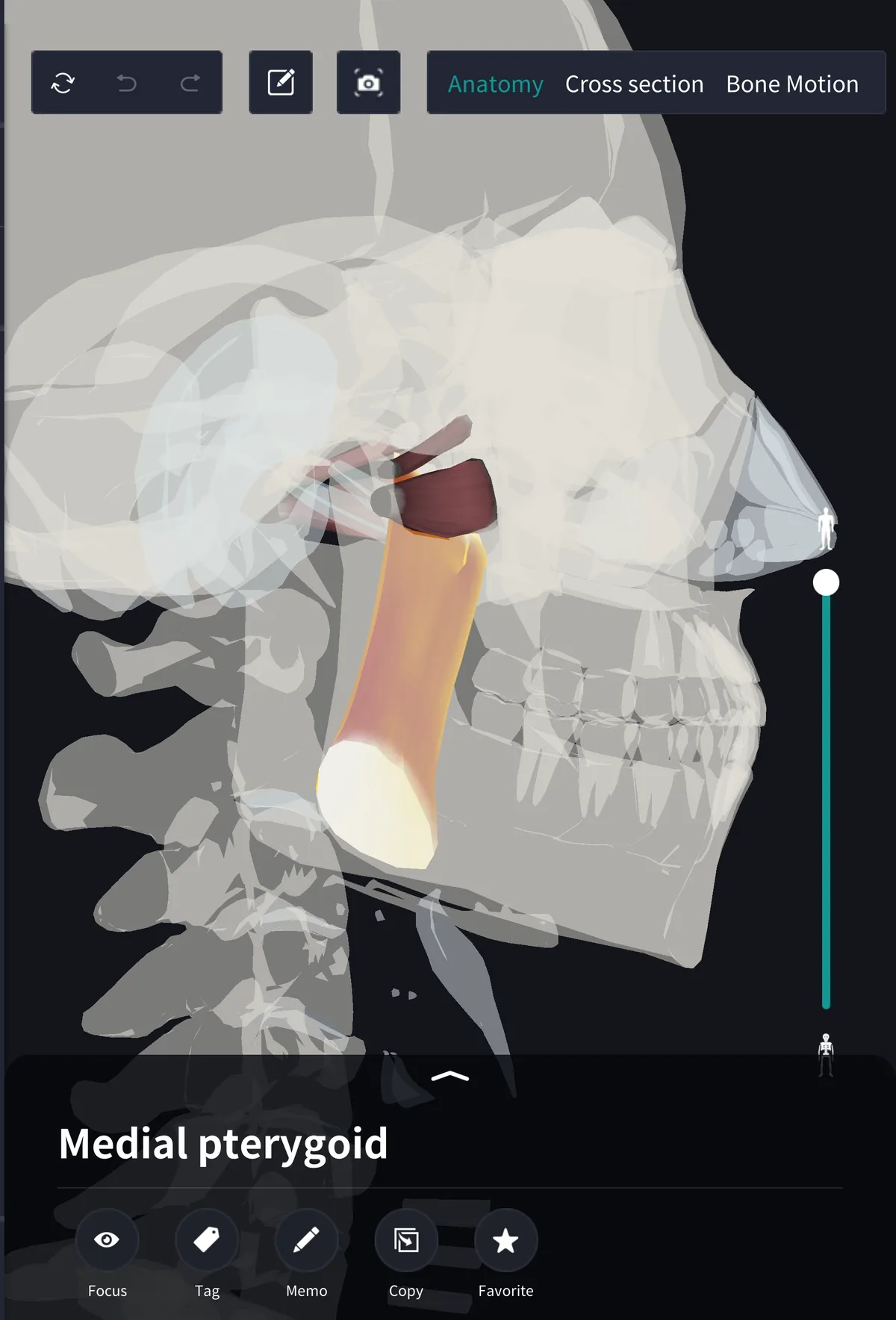
With the masseter and temporalis hidden, the two pterygoids are left: the medial runs vertically, the lateral almost horizontally. The masseter and medial pterygoid clasp the angle of the mandible from outside and inside, forming a sling that lifts the jaw.
Three close, one opens
The masseter, temporalis and medial pterygoid close the jaw. The lateral pterygoid runs almost horizontally and pulls the condylar process forward, protruding the jaw and starting wide opening. The posterior fibres of the temporalis pull a protruded jaw back. Grinding comes from the pterygoids of each side working in turn: when the medial and lateral pterygoids of one side contract together, the jaw shifts to the opposite side.
One nerve: the mandibular nerve
All four are supplied by branches of the mandibular nerve, the third division of the trigeminal nerve: the masseteric, deep temporal, and medial and lateral pterygoid nerves. The muscles of facial expression, such as the zygomaticus major and orbicularis oris, are supplied by the facial nerve instead.
Name and terms
Mastication means chewing; the group is also called the masticatory muscles. Pterygoid comes from the Greek for wing-like, after the pterygoid processes of the sphenoid bone they arise from.
See it in the app
The pterygoids lie inside the ramus of the mandible, out of reach of the fingers and hidden behind the masseter in side views. In the 3D anatomy app teamLabBody you can hide the masseter and temporalis, keep only the two pterygoids, and rotate them to compare their directions. The masseter is shown as separate superficial and deep parts. Cross sections are available with the PREMIUM plan.
FAQ
- How many muscles of mastication are there?
- Four: masseter, temporalis, medial pterygoid and lateral pterygoid. Some textbooks also count the digastric and other suprahyoid muscles as accessory muscles.
- Which muscle of mastication opens the mouth?
- The lateral pterygoid. It pulls the condylar process forward and helps start opening. The other three close the jaw.
- Which nerve supplies the muscles of mastication?
- All four are supplied by branches of the mandibular nerve, the third division of the trigeminal nerve.
Related articles
・Masseter: the muscle that hardens behind the cheek
・Superficial and deep masseter: the two layers
・Temporalis: the fan-shaped muscle at the temple
・Lateral and medial pterygoid: the muscles inside the ramus
・Zygomatic bone: forms the arch the masseter arises from